


















Lesson 4 of 45 · Getting Started
Stains, Culture Media and Aseptic Technique
Learn how the Gram stain and other stains work, how microbes are grown on agar, how to count colonies from serial dilutions, and the basics of lab safety.
Bacteria are nearly transparent, so under a bright-field microscope they look like faint ghosts. Stains solve the problem by coloring cells, and some stains do much more than that: they sort bacteria into groups that behave differently in the body and respond to different drugs. Once you can see microbes, the next challenge is to grow them, count them and keep them from escaping or being contaminated. That is the job of culture media and aseptic technique.
This lesson describes how these methods work. Everything about laboratory practice here is explained, not reported as an experiment of ours; real procedures must be done under supervision with your institution's safety rules.
What you will learn
- How a smear is prepared and why stains bind to cells.
- The steps of the Gram stain and what the result tells you about the cell wall.
- What acid-fast, endospore and negative stains are used for.
- The main types of culture media and how a streak plate isolates a colony.
- How to calculate CFU per milliliter from a dilution series.
- The basic safety levels used in microbiology laboratories.
Key terms
Smear: a thin film of cells dried onto a glass slide and heat-fixed so the cells stick.
Differential stain: a stain that makes different cell types look different.
Counterstain: a second dye that colors the cells that lost the first dye.
Colony: a visible mound of cells that all descend from one original cell or a few cells (a colony-forming unit, CFU).
Aseptic technique: methods that keep unwanted microbes out of a culture and protect the worker.
How stains work
Most dyes used in microbiology are charged molecules. Basic dyes such as crystal violet, methylene blue and safranin carry a positive charge. Bacterial surfaces are rich in negatively charged groups (phosphates and carboxyl groups), so basic dyes bind to the cells and color them. Acidic dyes such as nigrosin are negatively charged and are repelled by the cell surface, so they stain the background and leave cells as bright shapes. That is called a negative stain, and it is gentle enough to show the shape and capsule of cells without heating them, which can shrink them.
To prepare a standard stain, a drop of culture is spread on a slide, air-dried and passed through a flame (heat-fixing) so that the cells stay attached through the washes. A simple stain uses one dye to show size, shape and arrangement of cells. A differential stain uses several reagents to reveal differences between cells.
The Gram stain
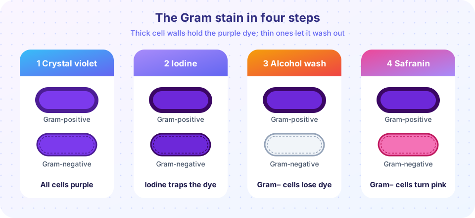
The Danish physician Hans Christian Gram devised this method in 1884, and it remains the first test done on many specimens. It has four steps:
- Crystal violet (the primary stain) colors all cells purple.
- Gram's iodine is a mordant: it forms a large complex with crystal violet inside the cell.
- Alcohol or acetone is the decolorizer. In cells with a thick peptidoglycan wall the solvent shrinks the pores and the complex stays trapped. In cells with a thin wall and an outer membrane the solvent dissolves the membrane lipids and the complex washes out.
- Safranin (the counterstain) colors the now colorless cells pink or red.
The result: Gram-positive bacteria stay purple, Gram-negative bacteria are pink. The difference reflects cell wall structure, which you will study in Chapter 3, and that structure predicts how bacteria respond to antibiotics. It also helps with identification: for example, Staphylococcus aureus is Gram-positive and appears as purple clusters of spheres, while Escherichia coli is Gram-negative and appears as pink rods.
Timing matters. If the alcohol wash is too long, even Gram-positive cells lose the dye and look pink; if too short, Gram-negative cells stay purple. Old cultures of some Gram-positive species can also stain as negative, so the stain is read from young cultures.
Other stains
- Acid-fast stain (Ziehl-Neelsen): hot carbolfuchsin penetrates waxy mycolic acids in the walls of Mycobacterium species, and acid-alcohol cannot remove it. Acid-fast cells stay red against a blue counterstain, which is how tuberculosis bacteria are detected in sputum smears.
- Endospore stain: heated malachite green enters the tough endospores of Bacillus and Clostridium, and safranin then colors the rest of the cell. Spores appear green inside or beside pink cells.
- Capsule stain: capsules are not stained directly; they show up as clear halos around cells against a dark background of a negative stain.
Culture media
A medium is a nutrient mixture. Liquid media are broths; solid media are made by adding agar, a gelling polysaccharide from red seaweed that melts near 85 °C but stays liquid only until it cools to roughly 40 °C, so it can be poured at about 50 °C and then sets into a firm gel at room temperature. Most microbes cannot digest agar.
| Medium type | Idea | Example |
|---|---|---|
| Complex | contains extracts of unknown exact composition | nutrient agar, tryptic soy agar |
| Defined (synthetic) | every ingredient and amount is known | glucose-salts medium |
| Selective | inhibits some microbes, allows others | MacConkey agar (bile salts favor Gram-negative rods) |
| Differential | colonies look different by a biochemical trait | MacConkey (lactose fermenters turn pink) |
| Enriched | extra nutrients for fussy species | blood agar |
Isolating and counting
To obtain a pure culture, a loop of a mixed sample is spread across an agar plate in a pattern (the streak plate), so that cells are separated far enough for one cell to grow into one colony. A colony is picked with a sterile loop and re-streaked to make sure it is pure.
To count how many living cells a sample contains, you make a serial dilution (for example, repeated tenfold dilutions), plate a known volume of several of them, and count the colonies on the plate that holds between 30 and 300. Fewer than 30 is statistically unreliable; more than 300 colonies crowd together. Then:
CFU per mL = colonies counted / (dilution factor × volume plated in mL)
Example: 87 colonies on a plate that received 0.1 mL of the 10⁻⁵ dilution gives 87 / (10⁻⁵ × 0.1) = 8.7 × 10⁷ CFU/mL in the original culture.
Press Run (or Ctrl + Enter) to see the result here.
Program input · what you'd type while the program runs
Type the answers before you press Run, one per line. Each time the program reads a line (with input()), it gets the next line from this box.
Aseptic technique and safety
Aseptic technique has a few habits that you will see in every teaching laboratory:
- Disinfect the bench before and after work, and wash hands.
- Flame an inoculating loop until red hot (or use a disposable loop), cool it, and flame the mouth of tubes.
- Keep plates closed, open them only as much as needed, and work close to a flame or in a biosafety cabinet.
- Never eat, drink or apply cosmetics in the laboratory, and never pipette by mouth.
- Sterilize waste, typically in an autoclave: steam under pressure at 121 °C for about 15 minutes or longer, which kills even endospores.
Laboratories are classified into four biosafety levels (BSL-1 to BSL-4) depending on the risk of the organisms handled. BSL-1 (teaching labs, harmless strains) needs standard practices; BSL-2 (most clinical labs) adds protective equipment and safety cabinets; BSL-3 handles serious airborne pathogens with controlled airflow; BSL-4 is for the most dangerous agents and uses full-body positive-pressure suits.
Common misconceptions
"Gram-positive means the bacteria are harmful (positive for disease)." The words refer only to staining. Both groups include harmless and dangerous species.
"Agar is a nutrient for the microbes." Agar is only the gel. The nutrients come from the broth that is added to it, which is why a plate of plain agar and water grows almost nothing.
"A colony is one cell." A colony is millions of cells, usually descended from one cell. That is why the unit is CFU, not "cells".
Check your understanding
Quick check · 5 questions
of 5 answered You scored / 5 · perfect!
-
1. In the Gram stain, which reagent is the decolorizer?
Alcohol washes the crystal violet-iodine complex out of cells with thin walls. Crystal violet is the primary stain, iodine the mordant and safranin the counterstain. -
2. A Gram-negative rod looks which color after a correct Gram stain?
It loses the purple complex in the alcohol step and then takes up the pink safranin. -
3. Why do acid-fast stains work on Mycobacterium?
Mycolic acids make the wall waxy; once hot carbolfuchsin enters, acid-alcohol cannot remove it. -
4. A plate that received 0.1 mL of a 10⁻⁴ dilution has 120 colonies. What is the CFU/mL of the original sample?
120 / (10⁻⁴ × 0.1) = 120 / 10⁻⁵ = 1.2 × 10⁷. -
5. Why is the 30 to 300 range used when counting colonies?
Small counts have large random variation, and crowded plates cannot be counted accurately.
Summary
- Basic dyes bind the negatively charged cell surface; acidic dyes give a negative stain.
- The Gram stain (crystal violet, iodine, alcohol, safranin) separates Gram-positive (purple) from Gram-negative (pink) bacteria by cell wall structure.
- Acid-fast and endospore stains detect special wall and spore structures.
- Agar plus a nutrient broth makes solid media; selective and differential media identify species. Streaking isolates colonies.
- CFU/mL = colonies / (dilution × volume plated); count plates with 30 to 300 colonies.
- Aseptic technique, autoclaving and biosafety levels protect the worker, the culture and the environment.
Track your progress
Create a free account to tick off lessons and pick up where you left off on any device.
of Microbiology lessons completed