


















Lesson 25 of 46 · T Cells and Cellular Immunity
MHC and the T cell receptor: how T cells see antigen
Learn how MHC class I and II present peptides, how the T cell receptor reads them, and why co-stimulation decides whether a T cell activates or shuts down.
A B cell receptor can grip a whole, folded molecule floating in the fluid. A T cell receptor is far pickier: it reads only a short peptide that is held in a groove of a cell-surface molecule from the major histocompatibility complex, or MHC. In humans these molecules are called HLA, for human leukocyte antigens. Two facts follow from this. Foreign proteins must be cut into peptides and loaded onto MHC before a T cell can notice them, and a T cell can only work with a partner cell that carries MHC.
This lesson covers how the loading works for the two classes of MHC, how a T cell receptor binds the result, and what has to happen next before a T cell will act. Understanding the requirement for a second signal is the key to understanding why the immune system usually does not attack your own tissues.
What you will learn
- How MHC class I and class II differ in location, peptide source and partner T cell.
- Why HLA genes are so variable and why that matters.
- The three signals needed to activate a naive T cell.
- What happens when signal 2 is missing.
Key terms
MHC / HLA: cell-surface proteins that hold peptides for T cells. HLA is the human name.
Antigen-presenting cell (APC): a cell that displays peptide on MHC class II and gives co-stimulation. Dendritic cells are the most powerful.
Co-stimulation: a second signal, usually CD28 on the T cell binding B7 (CD80 or CD86) on the APC.
MHC restriction: the rule that a T cell recognizes a peptide only when it is held by the MHC type the T cell was selected on.
Anergy: a state of long-lasting unresponsiveness in a T cell.
MHC class I: a display of what is inside
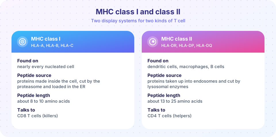
Almost every nucleated cell in the body carries MHC class I (the HLA-A, HLA-B and HLA-C genes). Class I molecules show peptides made from the cell's own proteins. The pathway is a sampling system:
- Proteins in the cell are continuously broken down by the proteasome into small peptides.
- A transporter called TAP moves peptides into the endoplasmic reticulum.
- There they are loaded onto new class I molecules, which are stabilized by a small partner protein, beta-2 microglobulin.
- The complex travels to the cell surface and holds its peptide, usually 8 to 10 amino acids long, on view.
This means a cell is constantly publishing a sample of everything it is making. A healthy cell displays self peptides, which T cells ignore. A virus-infected cell also displays peptides from viral proteins, and CD8 T cells that carry a matching receptor will recognize and kill it. The CD8 co-receptor binds to a constant part of class I, steadying the contact.
MHC class II: a display of what was eaten
MHC class II (HLA-DR, HLA-DP and HLA-DQ) is found mainly on professional antigen-presenting cells: dendritic cells, macrophages and B cells. These cells take in material from outside, in vesicles called endosomes. Lysosomal enzymes cut proteins into peptides, a protein called the invariant chain keeps the groove empty until class II reaches the right compartment, and the peptide is then loaded. Class II holds longer peptides, about 13 to 25 amino acids. The CD4 co-receptor binds class II, so CD4 T cells read class II and act as helpers.
Dendritic cells have a trick called cross-presentation: some can load peptides from ingested material onto class I as well. This allows them to activate CD8 T cells against viruses that never infected the dendritic cell itself.
Why HLA is so variable
The HLA genes are among the most variable genes in the human genome. Each gene exists in thousands of different versions (alleles) across the human population. Every person inherits one set of HLA genes from each parent, and both are expressed, which is called codominance. A person typically has up to six different class I molecules, and a similar number of class II molecules.
Why so much variation? Different HLA molecules bind different peptides, so a population with diverse HLA is harder for any one pathogen to evade completely. Individual variation also explains why transplants need matching, and why some HLA types are linked to particular diseases. The classic demonstration that T cells recognize peptide plus self-MHC together was made by Peter Doherty and Rolf Zinkernagel in 1974, work that earned them a Nobel Prize in 1996.
The T cell receptor and the synapse
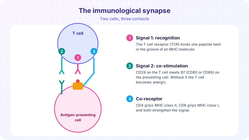
The T cell receptor is a heterodimer, a pair of chains (alpha and beta in most T cells), with a variable tip made of loops called complementarity-determining regions. These loops contact both the peptide and the walls of the MHC groove. The receptor itself has only a very short tail, so it is attached to a signaling complex called CD3, whose tails carry sequences that become phosphorylated when the receptor is engaged. The enzymes Lck and ZAP-70 carry the signal onward.
The contact between the T cell and the APC is a specialized zone called the immunological synapse. Receptors and signaling proteins gather there in a patterned arrangement, and the T cell may remain attached for hours to commit to activation.
The three signals
A naive T cell does not respond simply because it meets its peptide. It needs several signals together:
- Signal 1, recognition: TCR binds peptide-MHC, helped by CD4 or CD8.
- Signal 2, co-stimulation: CD28 on the T cell binds B7 molecules (CD80, CD86) on the APC. APCs only raise B7 when they have been alerted to danger by pattern recognition receptors, which is why this signal links innate and adaptive immunity.
- Signal 3, cytokines: signaling proteins released by the APC, such as IL-12, steer the T cell toward a particular effector type.
If a T cell gets signal 1 without signal 2, for example when it meets a self peptide on a calm tissue cell, it usually becomes anergic, or it is deleted. This is a major safety mechanism that guards against autoimmunity, and it is why vaccines include adjuvants, which supply the danger signals that raise co-stimulation.
Think of signal 1 as "which target?", signal 2 as "is this really an emergency?" and signal 3 as "what kind of response?".
Common misconceptions
"Every cell presents antigens to CD4 T cells." Only professional antigen-presenting cells carry MHC class II in large amounts. Most body cells carry class I only, and CD8 cells read those.
"Binding of the TCR is enough to activate a T cell." Binding alone gives signal 1 only. Without co-stimulation the cell can be silenced instead of activated.
"MHC molecules recognize antigens." MHC molecules merely hold peptides. The T cell receptor does the recognizing.
Try it
The first example is a decision function that applies the three-signal rule. It is a demonstration of the logic and not a model of the cell's real biochemistry.
Press Run (or Ctrl + Enter) to see the console output here.
No output.
The second example explores why variety in HLA helps. Suppose a given HLA molecule binds a random peptide with probability p. A person carrying more different alleles can present more of a pathogen's peptides. The value of p here is invented for illustration (real binding rates differ between alleles).
Press Run (or Ctrl + Enter) to see the result here.
Program input · what you'd type while the program runs
Type the answers before you press Run, one per line. Each time the program reads a line (with input()), it gets the next line from this box.
Check your understanding
Quick check · 5 questions
of 5 answered You scored / 5 · perfect!
-
1. Which cells normally carry MHC class II in large amounts?
Class II is mainly on dendritic cells, macrophages and B cells. Class I is on nearly every nucleated cell. -
2. Where do the peptides on MHC class I mainly come from?
The proteasome cuts cytosolic proteins and TAP carries the peptides to the ER for loading onto class I. -
3. A T cell receives signal 1 without signal 2. What is the most likely result?
Recognition without co-stimulation tends to silence the cell, a safeguard against autoimmunity. -
4. What does the CD4 co-receptor bind?
CD4 binds class II, so CD4 T cells read peptides on class II. CD8 binds class I. -
5. Why are adjuvants included in many vaccines?
Adjuvants activate innate sensors, which makes APCs express B7 and cytokines so that T cells respond rather than ignore the antigen.
Summary
- MHC class I presents peptides made inside cells (8-10 amino acids) to CD8 T cells. MHC class II presents peptides taken up from outside (13-25 amino acids) to CD4 T cells.
- HLA genes are highly variable and codominant, which broadens the range of peptides a person can present.
- The T cell receptor reads peptide plus MHC and signals through CD3, Lck and ZAP-70.
- Activation of a naive T cell requires three signals: recognition, co-stimulation (CD28-B7) and cytokines.
- Recognition without co-stimulation makes a T cell anergic, an important protection against self-attack.
Track your progress
Create a free account to tick off lessons and pick up where you left off on any device.
of Immunology lessons completed