


















Lesson 19 of 46 · Antigens and Antibodies
Antibody structure: heavy chains, light chains, Fab and Fc
Learn the Y-shaped structure of IgG: two heavy and two light chains, variable and constant domains, CDR loops, hinge, Fab and Fc fragments, and how structure explains function.
An antibody is one of the most carefully studied molecules in biology, and its shape explains nearly everything about what it does. At one end it carries two identical, adjustable grippers that fit an epitope. At the other end it carries a standard handle that tells other immune cells and proteins what to do next. In between is a flexible hinge. Variable grippers and a constant handle: that combination lets one design be used against millions of different targets while still triggering the same few effector systems.
In this lesson you will take an IgG antibody apart, domain by domain, learn the experiment that revealed its structure and see how the six loops at the tips decide what it binds.
What you will learn
- The four-chain structure of IgG and the names of its domains.
- Why the variable region differs between antibodies and the constant region does not.
- What CDRs are, and how they form the antigen-binding site.
- What Fab and Fc fragments are, and what each part does.
- The difference between affinity and avidity.
Key terms
- Immunoglobulin (Ig): the family of antibody molecules.
- Domain: a compact folded unit of about 110 amino acids (the immunoglobulin fold).
- Variable (V) / constant (C) region: the changing antigen-binding part and the fixed effector part.
- CDR: complementarity-determining region, a hypervariable loop that touches the antigen.
- Fab / Fc: the antigen-binding fragment (arm) and the crystallizable fragment (stem).
- Affinity / avidity: the strength of a single binding site versus the combined strength of several sites.
Four chains
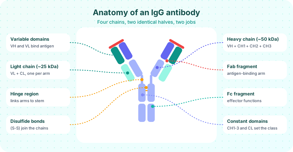
An IgG molecule has a total mass of about 150 kDa and is built from four polypeptide chains joined by disulfide bonds (covalent S-S bridges between cysteines) and by non-covalent contacts:
- Two identical heavy chains (H), each about 50 kDa (roughly 450 amino acids).
- Two identical light chains (L), each about 25 kDa (roughly 214 amino acids).
Each light chain is attached to one heavy chain, and the two heavy chains are joined to each other, giving the characteristic Y shape (sometimes drawn as a T because the hinge is flexible). Because the two halves are identical, an antibody has two identical antigen-binding sites; it is bivalent.
Each chain folds into domains, compact barrel-like units about 110 amino acids long made of two stacked β-sheets (the immunoglobulin fold). A light chain has two domains (VL and CL). An IgG heavy chain has four (VH, CH1, CH2, CH3). The antigen-binding site is formed by VH and VL together.
| Chain | Domains | Where they sit |
|---|---|---|
| Light | VL, CL | Outer side of each arm |
| Heavy | VH, CH1 | Arm (paired with the light chain) |
| Heavy | CH2, CH3 | Stem (Fc) |
| Between CH1 and CH2 | Hinge | Flexible linker |
Humans have two kinds of light chain, kappa (κ) and lambda (λ). Any one antibody has one or the other, never both; roughly 60 percent of human antibodies use kappa. There are five kinds of heavy chain, named by Greek letters: μ (mu), δ (delta), γ (gamma), α (alpha) and ε (epsilon), which define the classes IgM, IgD, IgG, IgA and IgE (next lesson).
Variable and constant regions
If you compare the amino acid sequences of many antibodies, the first ~110 amino acids of each chain (the V domains) are strikingly different from antibody to antibody. The rest (the C domains) are almost identical within a class. This observation, made in the 1960s, was a clue that antibody diversity came from the gene: a "variable" piece joined to a "constant" piece (confirmed in 1976 by Tonegawa).
Even within a V domain, the variability is concentrated in three short stretches called hypervariable regions or CDRs (CDR1, CDR2 and CDR3). They are loops at the tip of the domain. Between them lie more conserved framework regions that hold the scaffold. Six CDR loops (three from VH and three from VL) come together to form the antigen-binding surface, the paratope. CDR3 of the heavy chain is usually the most variable, because it lies at the junction where gene segments are joined (lesson 4).
Think of a hand: the framework is the palm and the CDRs are the fingertips. The palm is the same in every antibody, and the fingertips are adjusted to the target.
Fab and Fc
In the late 1950s Rodney Porter treated rabbit IgG with the enzyme papain. The enzyme cut the molecule at the hinge into three pieces of similar size:
- Two Fab fragments (fragment antigen-binding), each with one binding site (light chain plus VH and CH1).
- One Fc fragment (fragment crystallizable, because it crystallized easily), the two paired CH2 and CH3 domains.
Another enzyme, pepsin, cuts below the hinge disulfide bonds and yields one F(ab')₂ fragment (still bivalent) plus small pieces of the Fc. Both enzymes were crucial tools, and Porter and Gerald Edelman shared the 1972 Nobel Prize for working out antibody structure. Today, Fab fragments are used as medicines when the effector function of Fc is not wanted, for example in some treatments of the eye.
| Part | What it does |
|---|---|
| Fab (V domains, CDRs) | Binds the epitope; neutralizes toxins and viruses by blocking them |
| Hinge | Lets the arms swing and spread, so both can bind sites at various distances |
| Fc | Sets the "action": binds complement C1q, binds Fc receptors on phagocytes, NK cells and mast cells, and binds FcRn (neonatal Fc receptor) that carries IgG across the placenta and extends IgG half-life to about three weeks |
Affinity and avidity
Affinity is how tightly one binding site holds one epitope. It is usually expressed as the dissociation constant Kd: the concentration of antibody at which half of the epitopes are bound. A lower Kd means tighter binding. Typical antibodies after a mature response have Kd in the nanomolar range (10⁻⁹ M) and some reach picomolar (10⁻¹² M).
Avidity is the overall strength when several binding sites grip several epitopes at once. A bivalent IgG can attach with both arms to a surface covered in repeated epitopes and is much harder to dislodge than a single Fab. The pentameric IgM has ten sites, so even low-affinity IgM can have high avidity on a repetitive surface such as a bacterial coat.
Two forms of the same antibody
A B cell displays a membrane-bound copy of its antibody as the B-cell receptor (BCR). When the cell becomes a plasma cell, the heavy-chain mRNA is spliced differently so the tail that anchors it in the membrane is replaced by a short sequence that lets it be secreted. The antigen-binding specificity stays identical.
Common misconceptions
"Each antibody has one binding site." An IgG has two identical sites; IgM has ten; dimeric IgA has four.
"The whole antibody changes to fit a different target." Only the V domains (and mainly the six CDR loops) differ; the rest of the molecule is the same within a class.
"Light chains are just decoration." The light chain contributes three of the six CDR loops and affects binding.
Try it
Press Run (or Ctrl + Enter) to see the result here.
Program input · what you'd type while the program runs
Type the answers before you press Run, one per line. Each time the program reads a line (with input()), it gets the next line from this box.
Press Run (or Ctrl + Enter) to see the console output here.
No output.
Check your understanding
Quick check · 5 questions
of 5 answered You scored / 5 · perfect!
-
1. What is the approximate mass of an IgG molecule?
Two heavy chains (about 50 kDa each) plus two light chains (about 25 kDa each) give about 150 kDa. -
2. Which part of an antibody carries the CDR loops?
CDRs are hypervariable loops at the tips of VH and VL, forming the paratope. -
3. What did papain digestion of IgG produce?
Papain cuts at the hinge, separating the two arms from the stem. -
4. Which of these determines the class of an antibody (IgG, IgM and so on)?
The heavy chain type (gamma, mu, alpha, epsilon, delta) defines the class. -
5. What does a low Kd indicate?
Kd is the concentration that gives half-occupancy, so lower values mean tighter binding.
Summary
- IgG has two heavy and two light chains (about 150 kDa), built from immunoglobulin-fold domains and joined by disulfide bonds.
- The variable domains (VH, VL) contain six CDR loops that form the antigen-binding site; the constant region sets the class and effector functions.
- Papain splits IgG into two Fab arms and one Fc stem; pepsin gives F(ab')₂.
- Fc binds complement and Fc receptors; FcRn gives IgG its long half-life and placental transfer.
- Affinity is single-site strength (Kd); avidity is the combined strength of several sites.
Track your progress
Create a free account to tick off lessons and pick up where you left off on any device.
of Immunology lessons completed