


















Lesson 29 of 46 · Immune Regulation and Memory
Tolerance and regulatory T cells: how the immune system avoids self-attack
Understand central and peripheral tolerance, how Foxp3 regulatory T cells suppress responses, and how the CTLA-4 and PD-1 checkpoints keep T cells in check.
In the last chapter you saw that T cell receptors are built at random. Some of the cells that result will, by chance, recognize something that belongs to you. Others will make receptors that are fine in the thymus but meet a self antigen that the thymus never displayed. And any response to a real infection must eventually be turned down. All these problems fall under the heading of immunological tolerance and regulation, and the solution is not one mechanism but a stack of them.
The idea of tolerance has an interesting history. In 1945 Ray Owen noticed that twin calves that shared a placenta also shared blood cells without rejecting them. Peter Medawar later showed that mice exposed to foreign cells before birth tolerated grafts from the same donor in adulthood. Frank Macfarlane Burnet proposed that lymphocytes that react to self are eliminated during development, an idea that matured into the clonal selection theory. Medawar and Burnet shared the Nobel Prize in 1960.
What you will learn
- The difference between central and peripheral tolerance.
- How anergy, deletion and ignorance protect against self-reactive T cells.
- What regulatory T cells do and what happens when they are missing.
- How CTLA-4 and PD-1 act as checkpoints, and why they matter for cancer.
Key terms
Central tolerance: removal or editing of self-reactive lymphocytes in the thymus (T cells) or bone marrow (B cells).
Peripheral tolerance: mechanisms that act on mature lymphocytes in the tissues and lymph nodes.
Anergy: a functional shutdown of a lymphocyte that has met antigen without co-stimulation.
Foxp3: the master transcription factor of regulatory T cells.
Immune privilege: reduced immune activity at sites such as the eye, brain and testis.
Central tolerance
You studied the T cell version already: negative selection in the thymus removes thymocytes that bind self-peptide too strongly, and the AIRE protein lets the thymus show tissue-specific antigens. B cells have a similar process in the bone marrow. A B cell whose receptor binds a self antigen strongly may be deleted, may be given a second chance by receptor editing (rearranging the light chain again to change what the receptor recognizes), or may be silenced.
Central tolerance is effective but incomplete. Some self-reactive cells have a low affinity that escapes deletion, and some self antigens are never shown in the thymus or marrow, because they appear only late in life or only in a few places.
Peripheral tolerance
Cells that escape central tolerance face several more safeguards.
Ignorance. Self-reactive cells may simply never meet their antigen, for example because it is hidden behind a barrier or is at too low a concentration.
Anergy. As you saw in the last lesson, a T cell that gets signal 1 without co-stimulation becomes anergic. It does not divide or make IL-2 even if it later sees antigen properly.
Deletion. Strong repeated stimulation can make T cells display Fas and Fas ligand and commit activation-induced cell death. People with defects in Fas have a lymphoproliferative disease with autoimmune features.
Suppression. Regulatory T cells actively dampen their neighbours. This is the most powerful peripheral mechanism, and it is the subject of the next section.
Immune privilege. In the eye, the brain and the testis, barriers and local immunosuppressive signals restrict inflammation. The fetus in the womb is also protected by a combination of mechanisms at the placenta.
Regulatory T cells
Regulatory T cells, or Tregs, are CD4 T cells that carry the transcription factor Foxp3 and a high level of the IL-2 receptor alpha chain (CD25). In 1995 Shimon Sakaguchi and colleagues showed that removing CD25-positive CD4 T cells from mice caused them to develop widespread autoimmune disease, and that returning these cells prevented it. Later work identified Foxp3 as the key gene, building on the study of a spontaneous mouse mutant called scurfy and a human disease called IPEX (a severe, early-onset autoimmune condition due to mutations in the FOXP3 gene). Mary Brunkow, Fred Ramsdell and Sakaguchi were recognized with the 2025 Nobel Prize in Physiology or Medicine for discoveries concerning peripheral immune tolerance.
Tregs use several methods:
- Competing for IL-2. Tregs carry a lot of CD25 and absorb IL-2, starving other T cells of this growth factor.
- CTLA-4. This receptor on Tregs binds B7 on antigen-presenting cells more tightly than CD28 does, and it can remove B7 from the cell surface, reducing co-stimulation for everyone nearby.
- Suppressive cytokines. Tregs release IL-10, TGF-beta and IL-35, which dampen inflammation.
- Direct effects. Some Tregs can kill target cells with granzymes.
Two kinds are described: thymus-derived Tregs, which arise from strongly self-reactive thymocytes, and peripherally induced Tregs, which develop from naive T cells in tissues, particularly at the gut, in the presence of TGF-beta. The latter help tolerate harmless food antigens and gut bacteria.
Checkpoint receptors
Even activated effector T cells carry brakes. These are known as immune checkpoints.
- CTLA-4 is turned on soon after a T cell is activated and competes with CD28 for B7. It mostly limits the early priming phase in lymph nodes.
- PD-1 is a receptor that appears on activated and, later, exhausted T cells. Its partners, PD-L1 and PD-L2, are found on many tissue cells and on antigen-presenting cells. PD-1 signaling mostly limits effector T cells in the tissues themselves, protecting healthy tissue from collateral damage.
Chronic infection and cancer expose T cells to prolonged stimulation, and they gradually become exhausted, with high PD-1 and reduced function. Many tumors increase PD-L1 to switch off attacking T cells. James Allison, who studied CTLA-4, and Tasuku Honjo, who discovered PD-1, shared the 2018 Nobel Prize in Physiology or Medicine for showing that blocking these checkpoints can unleash an anti-tumor response. Lesson Cancer immunotherapy returns to this.
When regulation fails
| Failure | Consequence |
|---|---|
| No functioning Foxp3 (IPEX) | Severe multi-organ autoimmunity in infancy |
| No functioning AIRE | Autoimmune attack on endocrine glands and other organs |
| Defective Fas | Accumulation of lymphocytes, autoimmune features |
| Blocking CTLA-4 or PD-1 as therapy | Anti-tumor activity, but also immune-related side effects such as colitis |
These examples show that tolerance is active work. Even in a healthy person, self-reactive cells are present and held in check.
Common misconceptions
"Autoreactive cells are all deleted in the thymus." Healthy people carry self-reactive T and B cells. They are controlled by anergy, ignorance and regulatory cells.
"Tregs only suppress autoimmunity." They also limit responses to infection, which avoids tissue damage, and they help tolerate food and gut bacteria.
"Checkpoint receptors are defects." They are normal, essential brakes. Cancer exploits them, and checkpoint drugs release them on purpose.
Try it
The first model is a cascade filter. Starting with 1,000 self-reactive cells, each layer removes a fraction. The fractions are invented, but the lesson holds: independent layers multiply their effects.
Press Run (or Ctrl + Enter) to see the console output here.
No output.
The second model is a feedback loop with and without regulatory T cells. Effector cells grow when antigen is present. Tregs grow in proportion and suppress effectors. The equations are a toy to show behaviour, not a published model, and every parameter is invented.
Press Run (or Ctrl + Enter) to see the result here.
Program input · what you'd type while the program runs
Type the answers before you press Run, one per line. Each time the program reads a line (with input()), it gets the next line from this box.
Check your understanding
Quick check · 5 questions
of 5 answered You scored / 5 · perfect!
-
1. Which transcription factor defines regulatory T cells?
Foxp3 is the master regulator of Tregs, and mutations in FOXP3 cause IPEX. -
2. What is anergy?
Anergic cells are alive but unresponsive. -
3. Which checkpoint mainly limits effector T cells in tissues?
PD-1 acts mostly in peripheral tissues, while CTLA-4 acts early in lymph nodes. -
4. How do Tregs use CTLA-4?
CTLA-4 outcompetes CD28 for B7 and can strip B7 from antigen-presenting cells. -
5. Which pair of scientists shared the 2018 Nobel Prize for checkpoint therapy?
James Allison (CTLA-4) and Tasuku Honjo (PD-1) were honored in 2018.
Summary
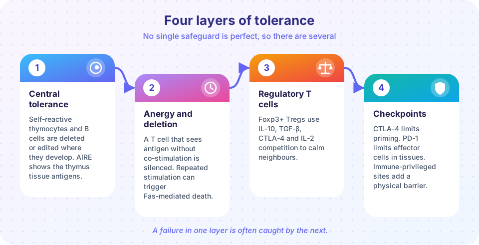
- Tolerance operates in layers: central deletion and editing, then ignorance, anergy, deletion, suppression and immune privilege in the periphery.
- Foxp3-positive regulatory T cells suppress other cells by IL-2 competition, CTLA-4, IL-10 and TGF-beta.
- CTLA-4 and PD-1 are checkpoints that limit T cell activity. Tumors exploit them, and drugs can block them.
- Failures of tolerance cause diseases such as IPEX and lead to autoimmunity.
Next: Immune memory
Track your progress
Create a free account to tick off lessons and pick up where you left off on any device.
of Immunology lessons completed