


















Lesson 9 of 46 · Overview of the Immune System
The lymphatic system and lymph nodes
Follow lymph from tissue fluid to lymphatic vessels, through lymph nodes and back to the blood; see the anatomy of a node and why nodes swell during infection.
Blood is not the only fluid that circulates. Alongside the blood vessels runs a second network, the lymphatic system, which does two jobs at once: it drains excess fluid from the tissues back into the bloodstream, and it carries antigens and immune cells to the places where the adaptive response begins. Anyone who has felt swollen glands in the neck during a sore throat has met this system in person.
In this lesson you will follow a droplet of fluid from the tissue to a node and back to the blood, learn the architecture of a lymph node, and understand why that architecture is so well suited to starting an immune response.
What you will learn
- Why lymph exists and where it flows.
- The main zones of a lymph node and which cells live in each.
- How lymphocytes enter and leave nodes.
- Why nodes swell, and what lymphatics have to do with cancer spread.
Key terms
- Lymph: the clear fluid collected from tissues and carried in lymphatic vessels.
- Afferent / efferent vessels: lymphatic vessels that bring lymph into a node (afferent) or carry it out (efferent).
- Germinal center: a region inside a B-cell follicle where B cells multiply and improve their antibodies.
- High endothelial venule (HEV): a special small blood vessel in nodes where lymphocytes leave the blood.
- Sentinel node: the first lymph node that drains a particular tissue.
Why lymph exists
Blood pressure pushes fluid out of the smallest blood vessels (capillaries) into the spaces between cells. Most of this fluid is taken back up by the capillaries, but not all of it. Textbook estimates are that around 20 liters leave the capillaries each day and about 17 liters are reabsorbed, leaving roughly 3 liters to return another way: through lymphatic vessels. Without them, fluid would accumulate and tissues would swell, a condition called lymphedema.
Lymph is similar to plasma but with less protein, and it carries extras: cells, cell debris, fats absorbed in the gut (milky lymph from the intestine is called chyle) and, crucially, antigens.
The route of lymph
- Initial lymphatics. Tiny blind-ended tubes in the tissue, with walls made of overlapping cells that act as one-way flaps. Pressure in the tissue pushes the flaps open and fluid enters.
- Collecting vessels. Larger vessels with valves that prevent backflow. The fluid is pushed along by the squeezing of nearby muscles, by breathing and by the rhythmic contraction of the vessel walls. There is no heart pumping lymph; that is why moving your legs helps swelling go down.
- Lymph nodes. On the way, lymph passes through one or more nodes, where it is filtered and inspected.
- Lymphatic trunks and ducts. The thoracic duct drains the left side of the body and everything below the diaphragm; the right lymphatic duct drains the right arm, right side of the head and right chest.
- Return to blood. Both ducts empty into large veins in the neck, at the junction of the subclavian and internal jugular veins.
Along the whole path lymph moves in only one direction: toward the heart.
Inside a lymph node
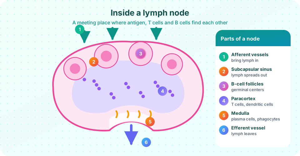
A node is an organized filter about the size of a bean (a few millimeters to a couple of centimeters). It has a fibrous capsule, and the cells inside are arranged in zones, as the picture shows.
- Afferent lymphatic vessels pierce the capsule and open into the subcapsular sinus, a channel under the capsule. Macrophages lining the sinus catch particles and pass antigens to B cells.
- The cortex holds B-cell follicles. In an active node some follicles contain a germinal center, where B cells divide rapidly, mutate their receptor genes and are selected for better antigen binding (Chapter 4).
- The paracortex lies deeper and is the T-cell zone. Dendritic cells that arrived from the tissues display antigen here to naive T cells.
- The medulla at the center and near the exit contains medullary cords, where plasma cells (antibody-secreting B cells) and macrophages sit. Antibodies leave the node with the efferent lymph.
- The efferent vessel exits at the hilum, the notch of the bean, where the node's artery and vein also pass.
The result: antigen enters at the edge, B cells see it first in the outer zones, T cells meet dendritic cells in the middle, and the products (antibodies and effector cells) exit through the medulla.
How lymphocytes find their way
Naive lymphocytes are constantly on the move, passing through node after node. They do not enter through the lymph; they leave the blood through high endothelial venules in the paracortex. Surface molecules such as L-selectin and the chemokine receptor CCR7 act like a passport and a signpost: chemokines made in the T zone attract T cells, while a different chemokine (CXCL13) draws B cells to follicles. A naive lymphocyte spends roughly a day in a node. If it finds no antigen it leaves in the efferent lymph and returns to the blood to try the next one. If it does find antigen, it stays, divides and sends its offspring out into the body.
This "recirculation" solves the needle-in-a-haystack problem: the rare lymphocyte that fits an antigen will sooner or later pass through the one node that is draining the infected tissue, because every node samples what drains from its own territory.
Why a node swells
During infection, a draining node enlarges as lymphocytes multiply, more blood flows in, and more cells are trapped inside. This reactive lymphadenopathy is typically tender and shrinks back after the infection is cleared. Nodes that become large, hard, painless or persistent need a doctor's evaluation. These notes are educational and are not a way to diagnose yourself.
Lymphatics also provide a route for metastasis: cancer cells can break off a tumor, travel in lymph and settle in the first node downstream. Surgeons therefore check the sentinel node to see whether a cancer has spread.
Common misconceptions
"Swollen glands mean something serious." Reactive nodes during colds and sore throats are common and normal. Persistent or unusual swelling should be assessed by a professional.
"Lymph is made by the nodes." Lymph comes from tissue fluid; nodes filter and change it.
"Lymphocytes get into nodes through lymph vessels." Naive lymphocytes enter nodes from the blood through HEVs; dendritic cells and antigen arrive via the lymph.
Try it
Press Run (or Ctrl + Enter) to see the console output here.
No output.
Press Run (or Ctrl + Enter) to see the result here.
Program input · what you'd type while the program runs
Type the answers before you press Run, one per line. Each time the program reads a line (with input()), it gets the next line from this box.
Check your understanding
Quick check · 5 questions
of 5 answered You scored / 5 · perfect!
-
1. Where do naive lymphocytes enter a lymph node?
Naive lymphocytes leave the blood at HEVs in the paracortex; antigen and dendritic cells arrive in afferent lymph. -
2. Which zone of a lymph node is mostly T cells and dendritic cells?
The paracortex is the T-cell zone where dendritic cells present antigen to naive T cells. -
3. What drives lymph forward through the vessels?
Lymph has no pump; valves ensure it moves only toward the heart as nearby muscles squeeze the vessels. -
4. About how much fluid returns to the blood as lymph each day, according to the textbook estimate used here?
Around 20 L are filtered and 17 L reabsorbed, leaving roughly 3 L for the lymphatics. -
5. Why do surgeons examine the sentinel node in some cancers?
Cancer cells that travel in lymph usually land in the first downstream node.
Summary
- The lymphatic system returns about 3 liters of fluid per day to the blood and carries antigen to nodes.
- Lymph flows one way through valved vessels, nodes and the thoracic and right lymphatic ducts into veins at the base of the neck.
- A node has a subcapsular sinus, B-cell follicles with germinal centers, a T-cell paracortex, a medulla with plasma cells and an efferent exit.
- Naive lymphocytes recirculate from blood through nodes, which solves the problem of finding a rare matching cell.
- Nodes swell during infection; lymphatics can also spread cancer.
Next: Chapter 2 review
Track your progress
Create a free account to tick off lessons and pick up where you left off on any device.
of Immunology lessons completed