


















Lesson 26 of 46 · T Cells and Cellular Immunity
Helper and killer T cells: the effector response
See how activated T cells expand into clones, how helper subsets Th1, Th2, Th17 and Tfh split the work, and how cytotoxic T cells kill infected cells with perforin and granzymes.
A naive T cell is a rare find. For any given peptide, only a handful of cells in your body carry a matching receptor. If the immune system waited for those few to do the whole job, an infection would win easily. The solution is clonal expansion: once a T cell is activated, it divides again and again, and its descendants all carry the same receptor. A single cell becomes a clone of thousands or more in about a week.
The offspring do not all behave the same way. CD4 cells differentiate into specialized helper subsets that direct the response. CD8 cells become cytotoxic effectors that destroy infected cells. This lesson covers both groups, the signals that choose between them, and what happens when the infection is over.
What you will learn
- How clonal expansion and contraction shape the size of a T cell response.
- The four major CD4 subsets, their cytokines and their targets.
- Why T follicular helper cells are essential for good antibody responses.
- How a cytotoxic T cell kills a target using perforin, granzymes and Fas.
Key terms
Clonal expansion: rapid division of an activated lymphocyte into many identical cells.
Cytokine: a small signaling protein released by cells to influence other cells.
Differentiation: a change of a cell into a specialized type with a specific job.
Perforin and granzymes: proteins from cytotoxic granules that open pores in a target cell and trigger its death.
Apoptosis: programmed cell death in which the cell dismantles itself in an orderly way.
Expansion, effector phase and contraction
After signals 1, 2 and 3, an activated T cell starts to divide, often every 6 to 12 hours. Interleukin-2 (IL-2), a growth factor that the activated cell both makes and responds to, drives much of this. In an acute viral infection, the number of antigen-specific CD8 T cells can rise thousands of times in a week or so, and they leave the lymph node and travel to the infected tissue.
When the infection is cleared, the stimulus disappears and most of the effector cells die by apoptosis, in a phase called contraction. Typically around 90 percent or more are removed. The survivors become memory cells, which you will study in Chapter 6. The three phases together, a surge, a fight and a cleanup, keep the response proportional to the threat.
Helper T cell subsets
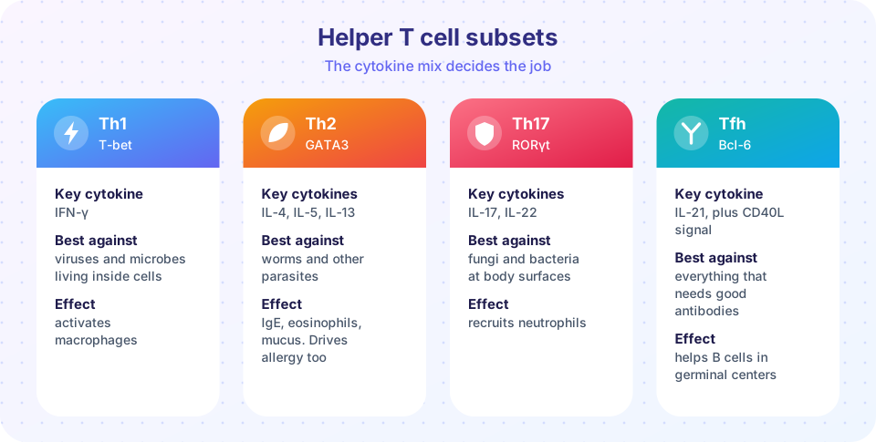
CD4 T cells do not kill anything themselves. They make decisions, mostly by releasing cytokines. Early studies in the 1980s by Tim Mosmann and Robert Coffman first showed that there were at least two types of helper cells, which they named Th1 and Th2. Since then more have been added. The cytokines present when a naive CD4 cell is activated decide which one forms.
Th1 cells appear in response to IL-12 and make interferon-gamma (IFN-gamma). Their master transcription factor is T-bet. IFN-gamma makes macrophages stronger killers, which is essential against microbes that survive inside macrophages, such as the tuberculosis bacterium. Defects in this pathway cause severe mycobacterial infections.
Th2 cells form in the presence of IL-4. Their transcription factor is GATA3, and they secrete IL-4, IL-5 and IL-13. These cytokines promote IgE, recruit eosinophils and increase mucus. That is the right response to large parasites such as intestinal worms, but the same machinery drives allergic disease when aimed at harmless antigens.
Th17 cells develop with TGF-beta and IL-6 (and related cytokines) and use the transcription factor ROR-gamma-t. They release IL-17 and IL-22 and recruit neutrophils, which fight extracellular bacteria and fungi at the skin and mucosal surfaces. Excessive Th17 activity is implicated in several autoimmune diseases, including psoriasis.
T follicular helper (Tfh) cells move into the B cell follicles of lymph nodes and spleen. They use the transcription factor Bcl-6 and the chemokine receptor CXCR5, and they provide IL-21 and a contact signal through CD40 ligand, which binds CD40 on B cells. This help is essential for germinal centers, where B cells improve their antibodies and switch from IgM to other classes. People who lack functional CD40 ligand have a condition with very low IgG, IgA and IgE.
A handy memory aid: Th1 = intracellular microbes, Th2 = parasites and allergy, Th17 = fungi and surface bacteria, Tfh = antibodies. Real responses are mixtures, and subsets can show some flexibility.
Cytotoxic T cells: the killers
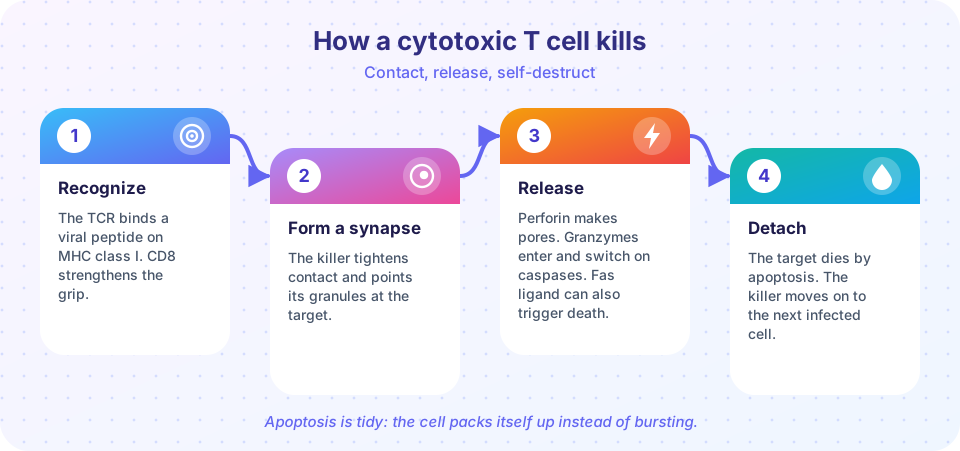
CD8 T cells that have been activated become cytotoxic T lymphocytes (CTLs). A CTL leaves the lymph node, circulates and checks other cells. When its receptor finds a matching peptide on MHC class I, it forms a tight synapse with the target and aims its cytotoxic granules at it. The granules contain two key ingredients:
- Perforin, which creates pores in the target's membrane.
- Granzymes, enzymes that enter through the damaged membrane and activate the target's own caspase enzymes, which dismantle the cell from within.
CTLs have a second route. The protein Fas ligand on their surface binds the Fas receptor on the target and sends a death signal. Whichever route is used, the target dies by apoptosis, not by bursting. This matters because apoptosis tidy-ly packages the cell's contents for disposal, limiting inflammation. It also destroys viral DNA or proteins in the dying cell, so any virus assembling inside is prevented from completing its production. A single CTL can kill several targets in succession, releasing each when it has done its work, and it also secretes IFN-gamma and tumor necrosis factor (TNF) to alert neighbours.
Natural killer (NK) cells use similar cytotoxic granules, but they are part of innate immunity and detect cells with low MHC class I rather than a specific peptide. T cell and NK cell killing complement each other: a virus that hides its MHC class I from CTLs becomes visible to NK cells.
The balance of help
CD8 responses to many infections depend on help from CD4 cells, because helper cells enhance the dendritic cell's ability to activate CD8 T cells and sustain memory. This is why HIV, which infects and destroys CD4 T cells, damages so many parts of the immune system and not only antibody production. A low CD4 count is the main laboratory marker of HIV progression.
Common misconceptions
"Th1 and Th2 are good and bad." Neither is better. Each is a tool. Th1 responses protect against intracellular microbes and Th2 against worms, but each causes disease when it is overactive or aimed at the wrong target.
"Killer T cells burst their targets open." They induce apoptosis. The target dies in an orderly fashion.
"Helper T cells are always CD4 and never kill." Almost all helpers are CD4, but rare CD4 cells can also have cytotoxic features. For learning purposes, think of CD4 as directing and CD8 as killing.
Try it
This model shows the surge-and-cleanup pattern with invented numbers. It grows a population of effector cells when antigen is present, removes about 90 percent when the antigen is gone, and keeps a few memory cells. Every number is illustrative.
Press Run (or Ctrl + Enter) to see the result here.
Program input · what you'd type while the program runs
Type the answers before you press Run, one per line. Each time the program reads a line (with input()), it gets the next line from this box.
This second example is a lookup table that maps a type of threat to the helper response that fits it best. It is a study aid, not a diagnostic tool.
Press Run (or Ctrl + Enter) to see the console output here.
No output.
Check your understanding
Quick check · 6 questions
of 6 answered You scored / 6 · perfect!
-
1. Which cytokine is the signature product of Th1 cells?
Th1 cells make interferon-gamma, which activates macrophages. -
2. Which cells give B cells the contact signal called CD40 ligand in germinal centers?
Tfh cells use CD40L and IL-21 to support B cells in the germinal center. -
3. How do cytotoxic T cells kill a target?
Perforin makes pores, granzymes activate caspases, and Fas ligand can trigger death through Fas. -
4. What usually happens to most effector T cells after an infection is cleared?
Contraction removes most effectors. A small fraction survives as memory cells. -
5. Which helper subset is best suited to fight intestinal worms?
Th2 cytokines promote IgE, eosinophils and mucus, the classic defence against parasites. -
6. Why does HIV infection weaken so many arms of immunity?
CD4 T cells direct B cells, CD8 cells and macrophages. Losing them has wide effects.
Summary
- Activated T cells divide rapidly into clones, act as effectors, and then mostly die in the contraction phase, leaving memory cells.
- CD4 helper subsets differ by cytokines and transcription factors: Th1 (T-bet, IFN-gamma), Th2 (GATA3, IL-4/5/13), Th17 (ROR-gamma-t, IL-17/22) and Tfh (Bcl-6, IL-21, CD40L).
- CD8 cytotoxic T cells recognize peptide on MHC class I and kill by perforin, granzymes and Fas ligand, causing apoptosis.
- CD4 help supports CD8 responses and antibody production, which is why losing CD4 cells is so damaging.
Next: Chapter 5 review
Track your progress
Create a free account to tick off lessons and pick up where you left off on any device.
of Immunology lessons completed