


















Lesson 7 of 46 · Overview of the Immune System
Lymphoid organs and the cells of immunity
Learn where immune cells are made and trained (bone marrow, thymus), where they meet antigen (nodes, spleen, MALT) and meet the family of white blood cells and their jobs.
Every immune cell is born somewhere, goes to school somewhere and goes to work somewhere. If you know those three places, the system stops being a bag of strange names and becomes a story with a plot. This lesson follows the story from the stem cell in the bone marrow to the lymphocyte waiting in a lymph node, and introduces the main cast of cells with one-line job descriptions.
What you will learn
- The difference between primary and secondary lymphoid organs.
- What the thymus, bone marrow, lymph nodes, spleen and mucosal tissues each do.
- The main branches of hematopoiesis.
- The job of each major white blood cell type.
Key terms
- Hematopoiesis: the production of all blood cells from stem cells.
- Primary lymphoid organ: an organ where lymphocytes develop (bone marrow, thymus).
- Secondary lymphoid organ: an organ where mature lymphocytes meet antigen (lymph nodes, spleen, MALT).
- Leukocyte: a white blood cell.
- MALT: mucosa-associated lymphoid tissue, found under the linings of the gut, airways and other mucosae.
Primary organs: the cell factory and the school
Bone marrow is the soft tissue inside bones such as the pelvis, sternum, ribs and vertebrae. Here hematopoietic stem cells (HSCs) divide to renew themselves and to produce progenitors that commit step by step to a lineage. A healthy adult makes on the order of hundreds of billions of blood cells a day, the vast majority red cells and neutrophils. The marrow is also where B cells develop and are tested; B stands for bursa of Fabricius (an organ in birds where they were first found) and conveniently also for bone marrow.
The thymus is a flat, two-lobed organ behind the breastbone. Its name gives T cells their letter. Immature T cells travel there from the marrow and spend days to weeks inside, where they are tested twice: first, can the T-cell receptor recognize our own antigen-presenting molecules at all (positive selection)? Second, does it bind too strongly to our own proteins (negative selection)? Cells that fail either test die. The large majority of developing T cells never graduate. The thymus is largest and most active in childhood and shrinks (involutes) with age, replaced partly by fat; adults still make T cells but more slowly, and long-lived memory cells carry much of the load.
You will meet selection in detail in Chapter 5. For now, remember the principle: lymphocytes are tested before they are released, to remove those that would attack the body.
Secondary organs: where meetings happen
A mature lymphocyte that has never met its antigen is called naive. It cannot be useful sitting in the marrow. It circulates through blood and lymph, visiting secondary organs, waiting for a match. These organs are designed to bring together antigen, antigen-presenting cells and lymphocytes in a small space.
- Lymph nodes (about 600 in the human body) filter lymph from the tissues. They are covered in the next lessons.
- The spleen filters blood. Its red pulp removes old red cells and traps microbes in the blood; its white pulp is a lymphoid area of T cells and B cells arranged around small arteries. People whose spleen has been removed or does not work are more at risk of serious infection with encapsulated bacteria and are advised to be vaccinated.
- MALT is a family of lymphoid tissues just under the surfaces that meet the outside world. It includes the tonsils and adenoids in the throat, Peyer's patches in the small intestine, and lymphoid tissue in the airways. By total cell number, the gut-associated part is the largest component of the immune system. Specialized M cells in the lining of Peyer's patches pass samples of gut contents to immune cells underneath.
The family tree of blood cells
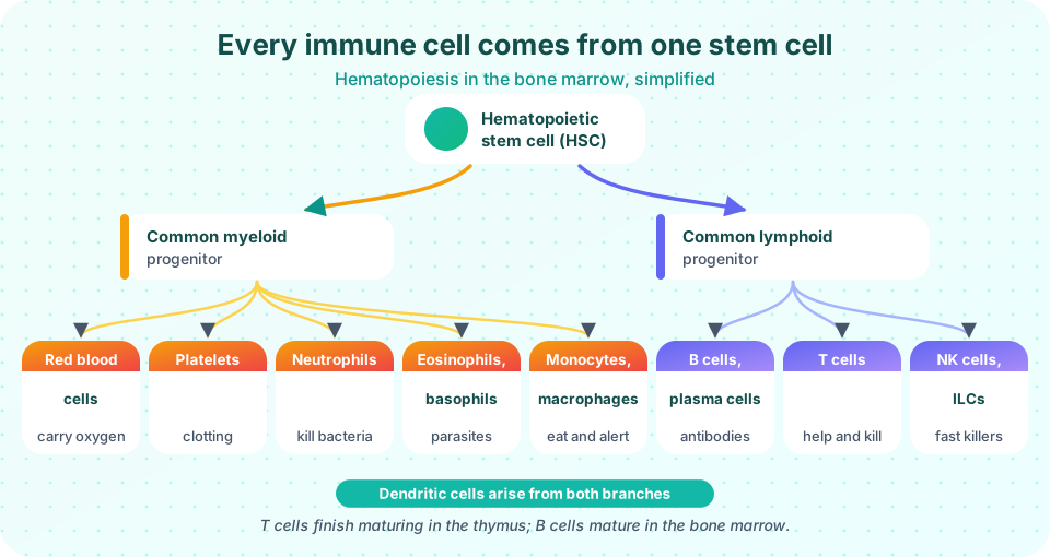
All blood cells descend from HSCs, as the picture shows. The first major split yields:
- The myeloid lineage: red blood cells (erythrocytes), platelets (from megakaryocytes), and the myeloid white cells: neutrophils, eosinophils, basophils, monocytes (which become macrophages in tissues), and most dendritic cells and mast cells.
- The lymphoid lineage: B cells, T cells, NK cells and innate lymphoid cells (ILCs).
Dendritic cells have more than one origin, which is why the picture shows them with a note rather than in a single box.
The cast of white blood cells
| Cell | Branch | Share of blood white cells (typical) | Main job |
|---|---|---|---|
| Neutrophil | Innate | 40 to 70% | First responder; engulfs and kills bacteria; lives only hours to a few days |
| Monocyte / macrophage | Innate | 2 to 8% (monocytes) | Eats microbes and debris; releases alarm signals; shows antigen to T cells |
| Dendritic cell | Bridge | Rare | Samples antigen in tissues, travels to nodes and activates naive T cells |
| Eosinophil | Innate | 1 to 4% | Attacks large parasites; involved in allergy |
| Basophil / mast cell | Innate | Under 1% (basophils) | Release histamine; allergy and parasite defense |
| NK cell | Innate | A fraction of lymphocytes | Kills virus-infected and abnormal cells without prior sensitization |
| B cell | Adaptive | Part of lymphocytes | Recognizes antigen; becomes an antibody-secreting plasma cell |
| T helper cell (CD4) | Adaptive | Part of lymphocytes | Coordinates other immune cells |
| T cytotoxic cell (CD8) | Adaptive | Part of lymphocytes | Kills infected cells |
CD stands for cluster of differentiation: a numbered system for labeling molecules on cell surfaces. CD4 and CD8 mark the two main kinds of T cell, and immunologists use these labels like name badges.
Granulocytes (neutrophils, eosinophils, basophils) are named for the granules in their cytoplasm that stain differently; mononuclear cells (monocytes and lymphocytes) have a single round nucleus.
A day in the life of a naive T cell
Imagine one naive T cell, born in the bone marrow, schooled in the thymus and released into the blood. Over the next day it leaves the blood through a high endothelial venule into a lymph node, spends hours in the T-cell zone pressing its receptors against the surface of dendritic cells, finds nothing that fits, and leaves through the efferent lymph. It is carried into the thoracic duct, back into the blood and on to the next node. It repeats this circuit many times, perhaps for months or years, until one day a dendritic cell carrying an infected finger's antigen presents a peptide that fits. Then the cell stops, divides and becomes an army. The organs in this lesson are simply the stations on that journey.
Common misconceptions
"White blood cells are all the same." There are at least a dozen distinct types with different jobs; a "high white count" can mean very different things depending on which type is high.
"Lymphocytes live in the blood." Most of the body's lymphocytes live in lymphoid tissues and only travel through the blood as a highway.
"The thymus disappears in adults." It shrinks and is partly replaced by fat but remains functional at a low level.
Try it
Press Run (or Ctrl + Enter) to see the result here.
Program input · what you'd type while the program runs
Type the answers before you press Run, one per line. Each time the program reads a line (with input()), it gets the next line from this box.
Check your understanding
Quick check · 5 questions
of 5 answered You scored / 5 · perfect!
-
1. Where do T cells complete their education?
Immature T cells leave the marrow and mature in the thymus, where positive and negative selection take place. -
2. Which pair are secondary lymphoid organs?
Secondary organs host the meeting of mature lymphocytes with antigen. -
3. Which cell is the most abundant white blood cell in a healthy adult?
Neutrophils make up roughly 40 to 70 percent of circulating white cells. -
4. What do CD4 and CD8 identify?
CD4 marks helper T cells and CD8 marks cytotoxic T cells. -
5. Which statement about the first split of hematopoiesis is correct?
The common myeloid and common lymphoid progenitors are the two main branches.
Summary
- Primary organs (bone marrow, thymus) make and train lymphocytes; secondary organs (nodes, spleen, MALT) are where they meet antigen.
- All blood cells come from hematopoietic stem cells; the first split is myeloid versus lymphoid.
- Neutrophils are the most common white cell; lymphocytes are B, T and NK cells; dendritic cells bridge innate and adaptive immunity.
- CD numbers are surface labels, and the thymus tests T cells before release.
Track your progress
Create a free account to tick off lessons and pick up where you left off on any device.
of Immunology lessons completed