


















Lesson 12 of 46 · Innate Immunity
Barriers and inflammation: the first response
Learn how skin, mucus, acid and the microbiota keep microbes out, and what happens when a barrier breaks: histamine, widened vessels, neutrophil recruitment and the signs of inflammation.
The best defense is a wall. Before any immune cell takes part, your body has already blocked, washed away or poisoned most microbes it meets. This lesson looks first at these barriers and then at what happens when a barrier is broken: the dramatic, local emergency response called inflammation. You will see why a splinter in your finger turns red, warm, swollen and sore, and why that is the body doing its job rather than failing at it.
What you will learn
- The physical, chemical and biological barriers that protect the body's surfaces.
- How tissue-resident cells detect a break-in and raise an alarm.
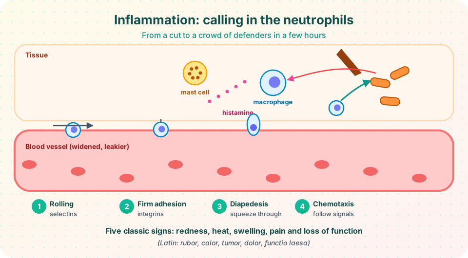
- The four steps by which neutrophils leave the blood and reach the wound.
- Why inflammation causes redness, heat, swelling and pain, and how it ends.
Key terms
- Epithelium: the sheet of cells covering a body surface or lining a cavity.
- Mucociliary escalator: mucus trapping particles in the airways while cilia sweep it upward.
- Microbiota: the community of microbes living on and in the body.
- Cytokine / chemokine: signaling proteins; chemokines specifically attract cells.
- Extravasation: the movement of cells from blood into tissue.
Layer 1 and 2: barriers
Skin. The outer layer (the stratum corneum) is made of dead, flattened, keratin-filled cells, packed tightly and constantly shed, which carries microbes away. Skin is dry, fairly acidic (about pH 5), salty from sweat and coated with oils (sebum) whose fatty acids hinder many microbes. Sweat and tears contain lysozyme, an enzyme that cuts the peptidoglycan wall of bacteria. Alexander Fleming discovered lysozyme in 1921 to 1922 (a few years before penicillin) when his own nasal mucus killed bacteria on a plate.
Mucous membranes. The linings of the airways, gut, urinary and reproductive tracts are thin, wet and more vulnerable, so they use other tricks. Goblet cells secrete mucus, a slimy gel of glycoproteins (mucins) that traps microbes. In the airways, cilia beat in coordinated waves and move the mucus up toward the throat to be swallowed or coughed out, the mucociliary escalator. Coughing, sneezing, vomiting, diarrhea and the flow of urine are mechanical flushing. Stomach acid (pH about 1 to 2) kills many swallowed microbes. Cells in the gut and skin also secrete defensins, short positively charged peptides that punch holes in microbial membranes.
The microbiota. Harmless bacteria cover your skin and fill your gut. They compete with invaders for space and food and release substances that inhibit newcomers, a phenomenon called colonization resistance. Broad-spectrum antibiotics can disturb this community and allow opportunists such as Clostridioides difficile to take over, which is one reason antibiotics are used only when needed.
When a barrier breaks
Suppose a splinter pierces the skin and carries bacteria into the tissue underneath.
- Detection. Resident macrophages and mast cells are sentinels. Their pattern-recognition receptors (next lesson) sense microbial molecules such as LPS or peptidoglycan, and also danger signals released by damaged cells.
- Alarm. They release cytokines (for example TNF-α, IL-1 and IL-6) and mediators such as histamine and prostaglandins.
- Vessel changes. The small blood vessels nearby widen (vasodilation), which increases blood flow, and their walls become leaky, letting plasma and proteins, including complement and antibodies, flow into the tissue.
- Recruitment. The cytokines make the vessel lining express adhesion molecules, so that passing neutrophils can stop and enter.
The four steps of neutrophil recruitment
| Step | What happens | Molecules involved |
|---|---|---|
| 1. Rolling | Neutrophils are slowed and roll along the vessel wall like a ball on Velcro | Selectins on the vessel lining and their sugar partners on neutrophils |
| 2. Firm adhesion | Signals switch the neutrophils' integrins to a sticky form, and the cells stop | Integrins (LFA-1) binding ICAM-1 on the lining |
| 3. Diapedesis | The cell squeezes between lining cells and through the vessel wall | PECAM-1 and others |
| 4. Chemotaxis | The neutrophil follows a chemical gradient to the source | Chemokines such as IL-8 (CXCL8), complement C5a, bacterial formyl peptides |
This sequence is the model for how almost every leukocyte, not only neutrophils, leaves the blood. When it fails because of inherited adhesion defects, patients suffer repeated infections with little pus, a striking proof that the steps matter.
The five signs of inflammation
The Roman writer Celsus described four signs in the first century, and the physician Galen added a fifth.
| Sign | Latin | Cause |
|---|---|---|
| Redness | rubor | More blood flows into the widened vessels |
| Heat | calor | Warm blood, plus metabolic activity |
| Swelling | tumor | Leaked plasma and cells collect in the tissue |
| Pain | dolor | Prostaglandins, bradykinin and swelling excite pain nerves |
| Loss of function | functio laesa | Pain and swelling discourage use of the area |
Systemic effects. A bigger infection spills cytokines into the blood. They act on the hypothalamus to raise body temperature (fever, via prostaglandin E2), prompt the liver to make acute-phase proteins such as C-reactive protein (CRP) and push the bone marrow to release more neutrophils. Doctors measure CRP in blood to gauge inflammation. Fever slows the growth of some microbes and speeds some immune processes, but it is also uncomfortable and sometimes dangerous, so persistent or very high fever needs medical attention.
How it ends
Neutrophils are short-lived and die at the site; the creamy material called pus is largely dead neutrophils and debris. Macrophages arrive later (hours to a day or two), clear the remains and switch to a repair mode that releases growth factors. The vessels return to normal and tissue heals, sometimes leaving a scar. If the stimulus does not go away, or the shut-off fails, inflammation becomes chronic and damages tissue. Many modern diseases (rheumatoid arthritis, atherosclerosis, inflammatory bowel disease) involve chronic inflammation.
Common anti-inflammatory drugs such as aspirin and ibuprofen reduce the production of prostaglandins by blocking the COX enzymes. This is an educational note; consult a pharmacist or doctor for medicines.
Common misconceptions
"Inflammation is a sign that something has gone wrong with the immune system." Acute inflammation is a protective, healthy response. Problems arise when it is excessive, misdirected or never switched off.
"Pus means the infection is getting worse." Pus is a sign that neutrophils have arrived and fought; its presence alone does not tell you whether the infection is winning or losing.
"A fever should always be lowered immediately." Fever is part of the defense. Whether to treat it depends on age, how high it is, how the person feels and other symptoms, so a clinician's advice is best.
Try it
Press Run (or Ctrl + Enter) to see the result here.
Program input · what you'd type while the program runs
Type the answers before you press Run, one per line. Each time the program reads a line (with input()), it gets the next line from this box.
Check your understanding
Quick check · 5 questions
of 5 answered You scored / 5 · perfect!
-
1. Which enzyme in tears and saliva cuts bacterial cell walls?
Lysozyme hydrolyzes peptidoglycan in bacterial walls, especially of Gram-positive bacteria. -
2. In neutrophil recruitment, which step involves integrins binding ICAM-1?
Rolling uses selectins; integrins on the neutrophil bind ICAM-1 on the vessel lining to stop the cell. -
3. What causes swelling (tumor) at a site of inflammation?
Increased vessel permeability lets plasma and proteins collect in the tissue. -
4. Which is NOT one of the five classic signs?
The five signs are rubor, calor, tumor, dolor and functio laesa. -
5. What is the main reason broad-spectrum antibiotics can allow infections such as C. difficile?
Losing normal gut bacteria removes colonization resistance.
Summary
- Skin, mucus, cilia, acid, lysozyme, defensins and the microbiota form the first two layers of defense.
- Tissue macrophages and mast cells detect a break-in, release cytokines and histamine and widen the vessels.
- Neutrophils reach the site by rolling, firm adhesion, diapedesis and chemotaxis.
- The five signs (redness, heat, swelling, pain, loss of function) come from vessel changes and mediators.
- Acute inflammation is protective and should resolve; chronic inflammation causes disease.
Track your progress
Create a free account to tick off lessons and pick up where you left off on any device.
of Immunology lessons completed