


















Lesson 27 of 46 · T Cells and Cellular Immunity
Chapter 5 review: T cells and cellular immunity
A one-page recap of T cell development, MHC presentation, the three activation signals and effector subsets, with a ten-question mixed quiz.
You have now followed a T cell from the thymus to the infected tissue. This review pulls the chapter into a single picture, a table you can use as a checklist, and a mixed quiz. If a row in the table feels fuzzy, go back to the lesson named beside it.
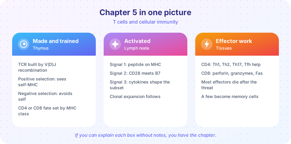
The chapter at a glance
| Topic | The key idea | Lesson |
|---|---|---|
| Receptor building | V(D)J recombination with RAG1 and RAG2 makes a random alpha-beta TCR. | Thymus |
| Positive selection | Cortex: keep cells that engage self-MHC. Decides CD4 versus CD8. | Thymus |
| Negative selection | Medulla: delete strongly self-reactive cells, with help from AIRE. | Thymus |
| MHC class I | On nearly all nucleated cells. Shows peptides made inside the cell to CD8 T cells. | MHC and TCR |
| MHC class II | On antigen-presenting cells. Shows peptides taken in from outside to CD4 T cells. | MHC and TCR |
| Three signals | Recognition, co-stimulation (CD28-B7), cytokines. Signal 1 alone leads to anergy. | MHC and TCR |
| Helper subsets | Th1, Th2, Th17 and Tfh, defined by transcription factor and cytokines. | Helper and killer |
| Killing | Perforin, granzymes and Fas ligand cause apoptosis. | Helper and killer |
| Expansion and contraction | A surge of division, then 90 percent or more die. Survivors form memory. | Helper and killer |
Ideas that connect
A few threads run through the whole chapter and are worth stating plainly.
Self is the reference point. The thymus does not test T cells against microbes. It tests them against self, and keeps the ones that are useful but not dangerous. Everything in tolerance, including the later chapter on regulation, builds on this.
MHC is the middleman. T cells cannot see free antigens. They see peptide-MHC complexes. The two MHC classes feed two T cell families and two defence tasks: class I leads to CD8 killers, class II leads to CD4 helpers.
Safety comes in layers. Selection in the thymus, the requirement for co-stimulation, and the contraction after an infection are three separate safety mechanisms. If one fails, another often catches the problem.
Context shapes the response. The same naive CD4 cell can become Th1, Th2, Th17 or Tfh depending on the cytokines present when it is activated. This is the link between innate and adaptive immunity.
Worked example: following one viral peptide
Pick a respiratory virus that infects the cells lining your airways. Trace one of its proteins through the chapter and you will touch every lesson.
Inside an infected epithelial cell, the viral protein is made on ribosomes and, like any protein, some of it is broken up by the proteasome. A few of the fragments are carried by TAP into the endoplasmic reticulum and loaded on MHC class I. The cell now displays a viral peptide at its surface, which is a warning flag. Meanwhile a dendritic cell in the tissue has picked up debris from dying infected cells. It matures, because pattern recognition receptors told it there is danger, and migrates to a lymph node carrying peptides on MHC class II, and on class I as well through cross-presentation.
In the lymph node, the dendritic cell meets a very large number of naive T cells. Only the few whose receptors, built by V(D)J recombination and passed by positive and negative selection, fit this peptide on this HLA type will bind. A CD4 cell gets signal 1 from the TCR, signal 2 from CD28 meeting B7 and signal 3 from IL-12, and it becomes a Th1 cell. A CD8 cell with a matching receptor receives the same signals, helped by the CD4 cell, and begins a burst of clonal expansion. A different CD4 cell becomes a Tfh cell and helps a B cell in a germinal center make better antibody.
Several days later the cytotoxic cells arrive in the airway. They find infected cells displaying the viral peptide on class I, form synapses and release perforin and granzymes. The cells die by apoptosis. When the virus is gone, most of the effector cells die as well, and a few remain as memory cells.
Notice what would break the chain. Without positive selection, no T cell would read the peptide. Without negative selection, some would attack the airway lining itself. Without co-stimulation, the dendritic cell could not activate anything. Without CD4 help, the killer response would be weaker and fade sooner. Every lesson of the chapter is one link in this chain.
Questions to ask yourself
- At which step does the cell make a decision between survival and death, and what information does it use?
- Which MHC class would a given cell use, and which co-receptor reads it?
- What is the difference between a signal that says what and a signal that says how much danger?
Try it
Use this short program to quiz yourself. It prints a prompt, and you can check each answer in your head before reading the next line. The pairs are drawn from this chapter.
Press Run (or Ctrl + Enter) to see the console output here.
No output.
The second activity checks the chapter's size logic. A beta chain with 45 V, 2 D and 13 J segments has this many simple V-D-J combinations (the segment counts are rounded teaching values).
Press Run (or Ctrl + Enter) to see the result here.
Program input · what you'd type while the program runs
Type the answers before you press Run, one per line. Each time the program reads a line (with input()), it gets the next line from this box.
Check your understanding
Quick check · 10 questions
of 10 answered You scored / 10 · perfect!
-
1. Which pair correctly matches an MHC class with the T cell family that reads it?
CD4 binds MHC class II and CD8 binds class I. -
2. A thymocyte whose receptor cannot bind self-MHC at all will:
No engagement means no survival signal in positive selection. -
3. What distinguishes negative from positive selection?
Positive selection keeps cells that can bind self-MHC. Negative selection removes the dangerously self-reactive ones. -
4. Which signal is delivered by CD28 binding to B7?
CD28-B7 is the classic co-stimulatory signal 2. -
5. A T cell meets its peptide on a resting tissue cell with no B7. The usual outcome is:
Signal 1 without signal 2 silences or removes the cell. -
6. Which helper subset supports germinal center B cells?
Tfh cells provide IL-21 and CD40L to B cells. -
7. Which two protein families make up the main killing weapon of a CTL?
Perforin forms pores and granzymes trigger apoptosis. -
8. What best describes the fate of most effector T cells after infection?
Contraction removes most effectors, leaving memory cells. -
9. Which protein lets thymic cells display peptides normally found in other organs?
AIRE switches on tissue-specific genes in thymic epithelial cells. -
10. Which statement about T cell receptors is correct?
The TCR reads peptide plus MHC. It is not secreted, and it does not bind whole microbes.
Summary
- T cells are made by random gene rearrangement, then tested against self in the thymus.
- They need peptide-MHC, co-stimulation and cytokines to become effectors.
- CD4 helpers split into Th1, Th2, Th17 and Tfh; CD8 cells kill with perforin, granzymes and Fas.
- Most effectors die after the infection, and a few remain as memory cells, which is the subject of the next chapter.
Track your progress
Create a free account to tick off lessons and pick up where you left off on any device.
of Immunology lessons completed