


















Lesson 30 of 46 · Immune Regulation and Memory
Immune memory: why the second encounter is faster and stronger
Compare primary and secondary immune responses, see how memory B cells, plasma cells and memory T cells are made, and learn how long protection can last.
Ancient historians noticed that people who survived plague or smallpox rarely caught it again. In 430 BCE, Thucydides wrote that those who had recovered from the plague in Athens could safely nurse the sick. Immunology gave the effect a name, immune memory, and found its cellular basis in lymphocytes that persist after an infection and respond faster and better when the same antigen returns.
Memory is what makes vaccines possible, and it is also why you have a good chance of getting measles only once. This lesson compares the first and second response in numbers, shows where memory is stored, and discusses how long it lasts.
What you will learn
- How the primary and secondary antibody responses differ in lag, size and antibody class.
- How germinal centers make memory B cells, plasma cells and higher-affinity antibodies.
- The main kinds of memory T cell and where they live.
- Why antibody levels fall but protection can last longer.
Key terms
Primary response: the response to a first encounter with an antigen.
Secondary response: the faster, stronger response on re-exposure.
Memory B cell: a long-lived B cell that can quickly become an antibody-secreting cell.
Long-lived plasma cell: an antibody-secreting cell that survives for years, often in the bone marrow.
Affinity maturation: improvement in antibody binding over time through mutation and selection.
Primary versus secondary response
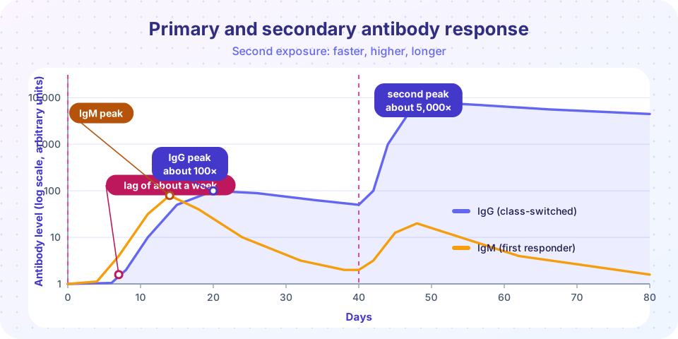
The first time your body meets an antigen, few lymphocytes recognize it, and they need time to find it, be activated and multiply. There is a delay of about 5 to 10 days before antibodies appear in blood. IgM, the first class made, appears first. Over the next weeks, B cells switch class, mainly to IgG, and the antibody levels reach a modest peak and then fall.
On re-exposure, many more cells are ready. The lag phase shrinks to about 1 to 3 days. The antibody level is much higher, often tens to hundreds of times, the antibody class is mostly IgG (or IgA and IgE at mucosal surfaces and in allergy), the antibodies bind more tightly, and the levels remain high for longer.
| Feature | Primary | Secondary |
|---|---|---|
| Lag before antibody appears | about 5-10 days | about 1-3 days |
| Peak level | modest | much higher |
| Main class | IgM first, then IgG | mostly IgG |
| Affinity | lower | higher (affinity maturation) |
| Duration | shorter | longer |
How B cell memory is made
When a B cell recognizes an antigen and receives help from a T follicular helper cell, it can enter a germinal center, a structure inside a lymph node follicle. There it goes through rounds of division. During this time, an enzyme called activation-induced cytidine deaminase (AID) introduces random point mutations in the antibody's variable genes, a process called somatic hypermutation. B cells whose mutated receptors bind antigen better are selected to survive and divide, and those that bind worse die. This cycle of mutation and selection is affinity maturation, and the same enzyme also drives class switching from IgM to IgG, IgA or IgE.
Germinal center B cells leave as two main products:
- Long-lived plasma cells that settle in niches, notably in the bone marrow, and secrete antibody continuously. They no longer divide and need no antigen to survive. They are the main source of the antibodies in your blood years after an infection.
- Memory B cells that circulate and wait. They do not secrete much antibody, but if the antigen returns they divide quickly and become plasma cells within days.
So there are two layers of B cell memory: a standing army of antibody that is already in the blood, and a reserve force that can be mobilized.
How T cell memory is made
After the contraction of an effector T cell response, a small share of the cells remain as memory T cells. Researchers distinguish several types, and the details are still being refined.
- Central memory (TCM) cells carry lymph node homing receptors such as CCR7 and CD62L. They circulate through lymphoid organs, divide a lot on re-exposure and generate new effectors.
- Effector memory (TEM) cells circulate through blood and tissues, and make effector molecules rapidly on encountering antigen.
- Tissue-resident memory (TRM) cells stay in the tissue where the infection happened, such as the skin, lungs or gut lining, and act as local sentries. Markers include CD69 and often CD103.
Memory T cells need less co-stimulation than naive cells, so they can respond to lower antigen doses and in less inflammatory settings. They are also more numerous than the original naive clone. They are kept alive by cytokines such as IL-7 and IL-15 and not by the antigen.
How long does memory last?
It depends on the pathogen, the vaccine and the person. Antibody levels fall over time because individual antibody molecules are broken down (IgG has a half-life of around three weeks in blood, thanks to a recycling receptor called FcRn) and because short-lived plasma cells die. Long-lived plasma cells keep a baseline going. In one well-known analysis, the levels of antibodies to measles, mumps and rubella were estimated to persist for decades, while antibodies to tetanus and diphtheria toxoids declined faster, with estimated half-lives of the order of a decade or two. These are estimates that depend on the method, so the exact figures should be treated cautiously.
Falling antibody levels do not always mean lost protection. Memory B and T cells can restart the response when exposed to the pathogen, which is why some diseases with a long incubation period are blocked even when antibody levels are low. This is also why booster doses are used for vaccines whose memory fades quickly, such as tetanus in adults and whooping cough vaccines. For some pathogens that mutate rapidly, such as influenza viruses, the issue is not fading memory but a changed target.
A related idea is trained immunity: after some infections or vaccines (BCG is the best-known example) innate cells such as monocytes may be reprogrammed to respond more strongly to later challenges. It is a different mechanism from classical memory and its practical importance is still under study.
Common misconceptions
"Memory means antibodies in the blood." Antibodies are one product. Memory B and T cells, which stay quiet until needed, are an equally important part.
"If antibody tests are negative, you are not immune." Cellular memory can remain even when antibody levels drop below a detection limit.
"Immunity is all or nothing." Memory reduces the chance and severity of disease, but it can be partial, and it can fade or be evaded by a mutating pathogen.
Try it
This model draws a primary and secondary response as simple curves with the lag times above. The heights, lags and decay rates are invented for illustration, not data from a real vaccine.
Press Run (or Ctrl + Enter) to see the result here.
Program input · what you'd type while the program runs
Type the answers before you press Run, one per line. Each time the program reads a line (with input()), it gets the next line from this box.
The second tool turns the half-life idea into a calculator: given a starting antibody level, a half-life and a protective threshold, how long until the level drops below it? The threshold and half-lives you try are your own assumptions. Real protective thresholds are specific to each pathogen and test.
Press Run (or Ctrl + Enter) to see the console output here.
No output.
Check your understanding
Quick check · 5 questions
of 5 answered You scored / 5 · perfect!
-
1. Which feature distinguishes the secondary response from the primary response?
Memory cells respond faster and produce more, higher-affinity IgG. -
2. Which enzyme drives somatic hypermutation and class switching in germinal centers?
Activation-induced cytidine deaminase (AID) introduces mutations in the antibody genes and enables class switching. -
3. Where do many long-lived plasma cells reside?
Bone marrow niches support long-lived plasma cells that secrete antibody continuously. -
4. Which memory T cell type stays in the tissue of the original infection?
TRM cells remain in tissues such as skin, lung and gut as local sentries. -
5. Why might a booster dose be useful?
A booster re-stimulates memory cells and raises antibody levels.
Summary
- The primary response has a lag of about a week and a modest IgM then IgG peak. The secondary response starts within 1-3 days and is much larger and of higher affinity.
- Germinal centers produce memory B cells and long-lived plasma cells through somatic hypermutation, selection and class switching.
- Memory T cells come as central memory, effector memory and tissue-resident memory.
- Antibody levels decline, but memory cells can restore protection, and boosters renew it.
Next: Chapter 6 review
Track your progress
Create a free account to tick off lessons and pick up where you left off on any device.
of Immunology lessons completed