


















Lesson 36 of 43 · Signaling and Regulation
Receptors and second messengers
Compare the four receptor families, follow the GPCR to G protein to cAMP to PKA pathway, learn how signals amplify and switch off, and see how toxins and drugs interfere.
A hormone molecule in the blood is just a messenger that cannot enter most cells. To act, it must bind a receptor protein, and that binding must be turned into something the cell can use. This lesson covers the four main families of receptors and follows the best-understood pathway in detail: epinephrine binding a G-protein-coupled receptor, making cAMP and switching on a kinase.
What you will learn
- Distinguish the four receptor families and the signals each one handles.
- Trace the GPCR, G protein, adenylyl cyclase, cAMP and PKA pathway.
- Explain amplification and the ways signals are switched off.
- Describe second messengers (cAMP, Ca²⁺, IP₃, DAG) and how toxins exploit G proteins.
Key terms
G protein: a heterotrimeric protein (α, β, γ) that is active when the α subunit holds GTP and inactive when it holds GDP.
Adenylyl cyclase: a membrane enzyme that converts ATP to cyclic AMP (cAMP).
Protein kinase A (PKA): an enzyme switched on by cAMP that phosphorylates target proteins.
Phosphodiesterase: an enzyme that breaks cAMP to AMP and so ends the signal.
Amplification: one activated molecule producing many activated molecules at the next step.
Two kinds of signal
Hydrophilic signals such as peptide hormones (insulin, glucagon) and epinephrine cannot cross the lipid bilayer, so they bind receptors on the cell surface. Lipophilic signals such as steroid and thyroid hormones diffuse through the membrane and bind intracellular receptors, which then act on DNA.
Four receptor families
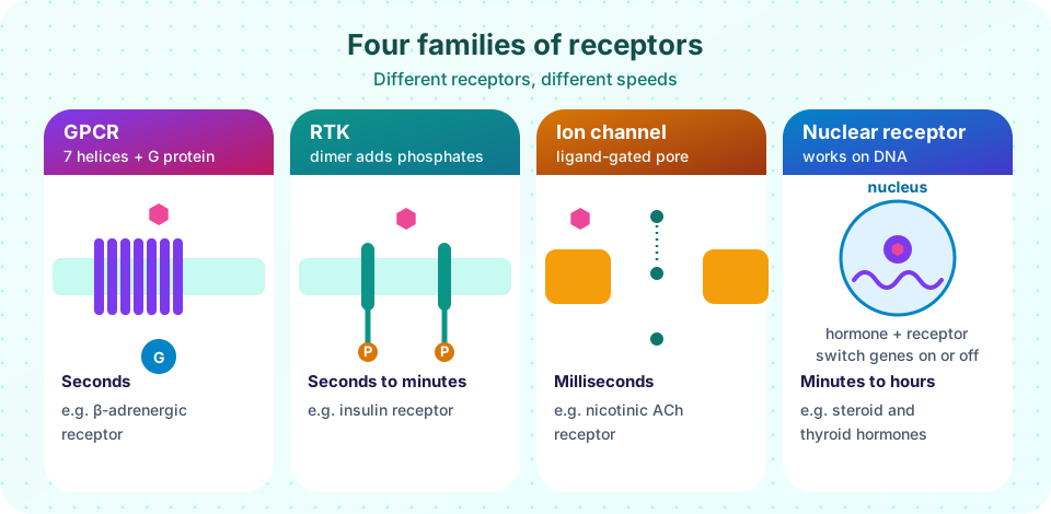
- G-protein-coupled receptors (GPCRs) have seven transmembrane α-helices and work through G proteins. They are the largest receptor family in the human genome and the target of a large share of drugs. Examples: β-adrenergic receptors, rhodopsin, many taste and olfactory receptors.
- Receptor tyrosine kinases (RTKs) have one helix and an intracellular kinase domain that adds phosphate to tyrosine residues on itself and on other proteins. Examples: the insulin receptor and the epidermal growth factor (EGF) receptor. Overactive RTKs are common in cancers.
- Ligand-gated ion channels open a pore when a ligand binds, changing ion flow in milliseconds. Example: the nicotinic acetylcholine receptor at the neuromuscular junction.
- Nuclear receptors bind steroid or thyroid hormones inside the cell and then bind DNA response elements to change gene expression over minutes to hours.
The GPCR to cAMP pathway
- Epinephrine binds a β-adrenergic receptor, changing its shape.
- The activated receptor acts as a guanine-nucleotide exchange factor for a G protein (Gs): the α subunit swaps GDP for GTP and separates from the βγ subunits.
- Gαs-GTP binds and activates adenylyl cyclase, which converts ATP into cAMP.
- cAMP binds the regulatory subunits of PKA, which release the catalytic subunits.
- PKA phosphorylates enzymes. In liver and muscle, this switches on glycogen breakdown (through phosphorylase kinase and glycogen phosphorylase, as in the polysaccharides lesson) and switches off glycogen synthesis.
Amplification. One activated receptor can activate many G proteins, each cyclase makes many cAMP and each PKA phosphorylates many enzymes. A few hormone molecules can therefore change the activity of many thousands of enzymes within seconds.
Switching off. Signals must stop. The α subunit has built-in GTPase activity: it hydrolyzes its GTP to GDP and returns to the inactive trimer, and RGS proteins speed this up. Phosphodiesterases break cAMP to AMP, phosphatases remove the phosphates, and the receptor itself is phosphorylated and removed from the surface.
Other second messengers
Gq-coupled receptors activate phospholipase C, which cuts a membrane lipid into IP₃ and diacylglycerol (DAG). IP₃ opens calcium channels in the endoplasmic reticulum; DAG and Ca²⁺ activate protein kinase C. Calcium is a second messenger because its resting concentration in the cytosol is very low (about 100 nM) compared with outside the cell (about 1 to 2 mM), so a small influx is a large relative change; calmodulin binds it and activates other enzymes.
RTKs work by phosphorylation: the phosphotyrosines are docking sites for adaptor proteins, which start the Ras–MAP kinase cascade (growth and division) or the PI3K–Akt pathway (survival and glucose uptake). The insulin receptor is already a dimer and changes shape on binding; EGF receptors dimerize when the ligand binds.
When signaling goes wrong
Cholera toxin modifies Gαs so that it cannot hydrolyze GTP; adenylyl cyclase stays on, cAMP rises in intestinal cells and the resulting salt and water loss causes severe diarrhea. Pertussis toxin modifies Gαi, which normally inhibits cyclase. Mutant, permanently active kinases (such as BCR-ABL in a type of leukemia) drive cells to divide, and drugs that block them were among the first targeted cancer therapies. This lesson is for learning mechanisms, not for medical advice.
Common misconceptions
"The hormone enters the cell." Peptide hormones and epinephrine stay outside; only the signal crosses.
"A signal stays on until the hormone leaves." Many controls act inside the cell: GTP hydrolysis, phosphodiesterases and phosphatases end the signal within seconds even if the hormone is still present.
"Second messengers are just waste products." cAMP, Ca²⁺ and IP₃ are made on purpose and removed on purpose.
Try it
How sensitive is the cell compared with the receptor? If a cell has spare receptors, the response can reach half of its maximum before half of the receptors are occupied. For a hyperbolic coupling with gain g, the half-maximal concentration is EC50 = Kd / (1 + g). The numbers are illustrative.
Press Run (or Ctrl + Enter) to see the result here.
Program input · what you'd type while the program runs
Type the answers before you press Run, one per line. Each time the program reads a line (with input()), it gets the next line from this box.
An amplification explorer. Each slider is how many molecules one molecule of the previous step activates (illustrative values, not measurements). The total is the product.
Check your understanding
Quick check · 5 questions
of 5 answered You scored / 5 · perfect!
-
1. Which receptor family opens in milliseconds?
Ligand-gated channels open a pore directly when a ligand binds; no cascade is needed. -
2. In the GPCR pathway, what makes cAMP?
Adenylyl cyclase, activated by Gαs-GTP, converts ATP to cAMP. -
3. How does a G protein switch itself off?
The α subunit has intrinsic GTPase activity that returns it to the inactive state. -
4. What does cholera toxin do?
By preventing GTP hydrolysis it keeps Gαs on and cAMP high, causing fluid loss from intestinal cells. -
5. Why can a cell respond to hormone concentrations below the receptor Kd?
With coupling gain, EC50 is lower than Kd because a small fraction of occupied receptors can generate a large response.
Summary
- Hydrophilic signals bind surface receptors; lipophilic signals act on intracellular receptors.
- The four receptor families are GPCRs, RTKs, ligand-gated channels and nuclear receptors.
- GPCR pathways use G proteins, adenylyl cyclase, cAMP and PKA, with strong amplification.
- Signals end by GTP hydrolysis, phosphodiesterases and phosphatases.
- Toxins and mutations that disrupt switches cause disease.
Track your progress
Create a free account to tick off lessons and pick up where you left off on any device.
of Biochemistry lessons completed