


















Lesson 32 of 43 · Nucleic Acids and Information Flow
DNA structure and replication
Learn nucleotide structure, Chargaff's rules, the antiparallel double helix, semi-conservative replication and the roles of helicase, primase, DNA polymerase and ligase.
DNA is a polymer with an astonishingly simple chemistry: four kinds of nucleotide in a long sequence. Its power comes from a structural trick. Each base can pair specifically with exactly one partner, so one strand contains all the information needed to rebuild the other. This lesson covers how nucleotides are joined, how the double helix is organized, and how the cell uses base pairing to copy its whole genome with very few mistakes.
What you will learn
- Describe the parts of a nucleotide and the phosphodiester bond.
- State Chargaff's rules and the features of the B-form double helix.
- Explain semi-conservative replication and the Meselson–Stahl experiment.
- Name the enzymes at the fork and explain leading and lagging strands.
- Estimate replication time from fork speed and genome size.
Key terms
Phosphodiester bond: the link between the 3′ hydroxyl of one nucleotide and the 5′ phosphate of the next.
Purine / pyrimidine: two-ring bases (A, G) / one-ring bases (C, T, U).
Semi-conservative replication: each new DNA duplex has one old strand and one new strand.
Okazaki fragment: a short piece of the lagging strand, about 100 to 200 nucleotides in eukaryotes and 1,000 to 2,000 in bacteria.
Origin of replication: the site where DNA unwinding and copying begin.
Nucleotides and the polymer
A nucleotide has a nitrogenous base, a five-carbon sugar and a phosphate group. In DNA the sugar is 2′-deoxyribose, which lacks the hydroxyl on carbon 2′. The bases are the purines adenine (A) and guanine (G) and the pyrimidines cytosine (C) and thymine (T). RNA uses ribose and replaces thymine by uracil (U).
Nucleotides are joined by phosphodiester bonds: the 3′ hydroxyl of one sugar attacks the α-phosphate of an incoming nucleoside triphosphate, releasing pyrophosphate, which is then hydrolyzed. The pyrophosphate hydrolysis makes the polymerization effectively irreversible. Each strand therefore has a free 5′ phosphate at one end and a free 3′ hydroxyl at the other, which gives it a direction.
The double helix
By 1950 Erwin Chargaff had shown that in DNA from many species the amount of A equals T and the amount of G equals C, while the A+T to G+C ratio differs from species to species. In 1953 James Watson and Francis Crick proposed a double helix, building on the X-ray diffraction work of Rosalind Franklin and Maurice Wilkins. Its features:
- Two antiparallel strands (5′→3′ and 3′→5′), twisted into a right-handed helix.
- Sugar-phosphate backbones on the outside, bases stacked inside.
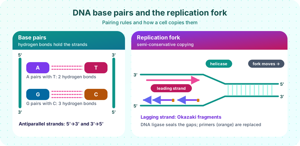
- A pairs with T by two hydrogen bonds and G pairs with C by three. A purine always pairs with a pyrimidine, so the width is constant, about 2 nm.
- In the common B form there are about 10 base pairs per turn, 0.34 nm per base pair and a major and a minor groove where proteins read the sequence.
The helix is stabilized more by base stacking (hydrophobic and van der Waals contacts between adjacent bases) than by hydrogen bonds alone. Because G–C pairs have three hydrogen bonds, DNA rich in G–C melts at a higher temperature. In cells DNA is also wound around histone proteins in nucleosomes (about 147 base pairs per nucleosome) and supercoiled by topoisomerases.
Semi-conservative replication
Watson and Crick noted that the structure suggested a copying mechanism. In 1958 Matthew Meselson and Franklin Stahl tested it by growing E. coli in heavy nitrogen (¹⁵N) and then switching to light nitrogen (¹⁴N). After one generation all DNA had intermediate density, and after two generations half was intermediate and half light, which ruled out the alternatives and confirmed that each daughter duplex keeps one old strand.
The replication fork
Replication starts at an origin, where proteins open the helix and form two replication forks moving in opposite directions.
- Helicase unwinds the duplex using ATP; single-strand binding proteins keep the strands apart; topoisomerase relieves the supercoiling ahead of the fork.
- Primase makes a short RNA primer, because DNA polymerase cannot start a chain from scratch; it can only extend a 3′ end.
- DNA polymerase (pol III in E. coli; pol δ and ε in eukaryotes) adds nucleotides to the 3′ end, always growing 5′→3′ and reading its template 3′→5′.
- On the leading strand the new DNA grows toward the fork, so it is made continuously. On the lagging strand the 5′→3′ rule means synthesis runs away from the fork, so the strand is made in pieces, the Okazaki fragments, each starting with its own primer.
- DNA polymerase I (bacteria) removes the RNA primers and fills the gaps, and DNA ligase seals the nick between fragments with ATP or NAD⁺ energy.
Accuracy is high because polymerases select the right nucleotide by shape, and many also proofread: a 3′→5′ exonuclease removes a mismatched base before extension continues. After mismatch repair the final error rate is roughly one in a billion bases or lower. Because the lagging strand's end cannot be primed, human chromosome ends shorten at each division unless telomerase, an enzyme carrying its own RNA template, extends them.
Common misconceptions
"Hydrogen bonds hold the helix together mainly." Base stacking contributes more stability; hydrogen bonds give specificity to pairing.
"The lagging strand is made backwards." It is made 5′→3′ like every strand; it is only discontinuous because that direction runs away from the fork.
"Replication copies one strand." Both strands are templates at the same time, at each fork.
Try it
A small sequence toolkit: reverse complement, G+C content and two melting temperature estimates (the quick Wallace rule for short oligonucleotides and a salt-adjusted formula for longer ones). The Tm numbers are estimates, not measurements.
Press Run (or Ctrl + Enter) to see the result here.
Program input · what you'd type while the program runs
Type the answers before you press Run, one per line. Each time the program reads a line (with input()), it gets the next line from this box.
Replication takes time. The numbers below are commonly quoted round figures: about 1,000 nucleotides per second per fork in E. coli and about 50 per second in human cells. The calculation shows why large genomes need many origins.
Press Run (or Ctrl + Enter) to see the result here.
Program input · what you'd type while the program runs
Type the answers before you press Run, one per line. Each time the program reads a line (with input()), it gets the next line from this box.
Check your understanding
Quick check · 5 questions
of 5 answered You scored / 5 · perfect!
-
1. Which base pair has three hydrogen bonds?
G–C pairs have three hydrogen bonds; A–T pairs have two. -
2. Why is the lagging strand made in fragments?
Because polymerases extend 3′ ends only, the strand template running the other way must be copied discontinuously. -
3. In the Meselson–Stahl experiment, after two generations in ¹⁴N the DNA was
Semi-conservative replication predicts two hybrid duplexes and two fully light duplexes after two rounds. -
4. What is the job of primase?
Polymerase cannot start a strand, so primase supplies an RNA primer. -
5. What gives DNA a higher melting temperature?
G–C pairs have three hydrogen bonds and stack strongly, so DNA rich in them melts at higher temperature.
Summary
- Nucleotides join by phosphodiester bonds; each strand has a 5′ and a 3′ end.
- Chargaff's rules and the antiparallel helix follow from A–T (two) and G–C (three) hydrogen-bonded pairs.
- Replication is semi-conservative; polymerases extend 3′ ends only, so the lagging strand is made in Okazaki fragments.
- Helicase, primase, polymerase, exonuclease proofreading and ligase work together at the fork.
- Large genomes need many origins to finish copying in a few hours.
Track your progress
Create a free account to tick off lessons and pick up where you left off on any device.
of Biochemistry lessons completed