


















Lesson 10 of 43 · Amino Acids and Proteins
Chapter 3 overview: amino acids and proteins
Overview of chapter 3: how twenty amino acids build proteins, the four levels of structure, folding, and how proteins such as hemoglobin bind other molecules.
If DNA is the recipe book of the cell, proteins are the cooks, the tools and much of the kitchen. Enzymes, antibodies, transporters, hormones such as insulin, structural fibers such as collagen and the motors of muscle are all proteins. They share a simple design, a chain of amino acids, yet their shapes and jobs are very different. This chapter explains how a sequence turns into a working machine.
What you will learn
- See how the lessons of chapter 3 build from residues to function.
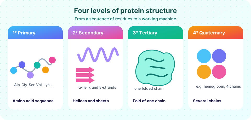
- Name the four levels of protein structure.
- Appreciate the size of sequence space and why sequence determines function.
The chapter at a glance
Twenty building blocks
The side chain decides the personality.
From chain to shape
Each level rests on the one below.
Folding
Misfolding causes disease.
Function
Structure changes allow regulation.
Where each lesson fits
- Amino acids: the general structure, the twenty standard side chains, ionization, zwitterions and the isoelectric point.
- The peptide bond and protein structure: the rigid peptide plane, α-helices, β-sheets and the four levels.
- Protein folding and stability: Anfinsen's experiment, the energy landscape, denaturation and misfolding.
- Protein binding and cooperativity: dissociation constants, myoglobin and hemoglobin, and the Hill equation.
- Chapter 3 review: a summary picture and ten questions.
Why do sequences matter so much?
A protein of 100 residues could in principle have any of 20¹⁰⁰ ≈ 1.3 × 10¹³⁰ sequences. Evolution has sampled an extremely small part of that space, and for most proteins even small changes can matter: a single amino acid change (glutamate to valine at position 6 of the β chain of hemoglobin) causes sickle cell disease. Yet many changes are tolerated, particularly on the surface, and proteins from different species often share the same fold with different sequences. Understanding which positions matter, and why, is at the heart of this chapter.
Common misconceptions
"A protein is a floppy string." Most proteins fold into a specific three-dimensional shape, which is essential for function. Some regions or whole proteins are intrinsically disordered, but that is an exception.
"One gene, one shape." Many proteins change shape when they bind a ligand, and many genes give more than one protein through splicing and modification.
Try it
First, a gallery of protein sizes. The sizes are approximate and use the average residue mass of about 110 Da to estimate the number of residues.
Press Run (or Ctrl + Enter) to see the result here.
Program input · what you'd type while the program runs
Type the answers before you press Run, one per line. Each time the program reads a line (with input()), it gets the next line from this box.
Second, a sequence-space calculator. It uses exact integer arithmetic to count the possible chains of a given length.
Press Run (or Ctrl + Enter) to see the console output here.
No output.
Check your understanding
Quick check · 4 questions
of 4 answered You scored / 4 · perfect!
-
1. Which is NOT a level of protein structure?
The standard levels are primary, secondary, tertiary and quaternary. -
2. How many possible sequences exist for a chain of 3 amino acids drawn from the 20 standard ones?
20 × 20 × 20 = 8,000. -
3. Sickle cell disease is caused by:
Glutamate at position 6 of the beta chain is replaced by valine, which makes deoxygenated hemoglobin stick together. -
4. The structure level that describes how several chains assemble is:
Quaternary structure is the arrangement of multiple polypeptide subunits.
Summary
- Proteins are chains of amino acids that fold into specific three-dimensional shapes.
- Four levels of structure: primary, secondary, tertiary and quaternary.
- Sequence determines structure, and structure determines function, but many substitutions are tolerated.
- This chapter moves from amino acids to folding and binding.
Next: Amino acids
Track your progress
Create a free account to tick off lessons and pick up where you left off on any device.
of Biochemistry lessons completed