


















Lesson 14 of 43 · Amino Acids and Proteins
Protein binding, myoglobin, hemoglobin and cooperativity
Learn how proteins bind ligands: dissociation constant Kd, saturation curves, why myoglobin stores oxygen and hemoglobin delivers it, and the Hill equation.
A protein is only useful if it can interact with something else: oxygen, a substrate, a hormone, DNA, another protein. The strength of that interaction, and how it responds to conditions, determines the protein's role. Two oxygen-binding proteins make a perfect case study. Myoglobin and hemoglobin bind the same ligand with the same kind of heme group, yet they behave very differently because one is a single unit and the other is a team.
What you will learn
- Define the dissociation constant Kd and the fractional saturation θ.
- Explain why myoglobin and hemoglobin have different curves.
- Use the Hill equation and interpret P50 and the Hill coefficient.
- Describe allosteric effectors such as H⁺, CO₂ and 2,3-bisphosphoglycerate (BPG).
Key terms
Ligand: a molecule that binds specifically to a protein.
Kd: the dissociation constant; the ligand concentration at which half the binding sites are occupied.
Fractional saturation (Y or θ): the fraction of binding sites occupied.
P50: the oxygen partial pressure at which hemoglobin or myoglobin is half saturated.
Cooperativity: binding at one site changes the affinity of other sites on the same protein.
Allosteric: regulated by binding at a site other than the active (or ligand) site.
Simple binding and Kd
For a protein P binding a ligand L at one site, P + L ⇌ PL, the dissociation constant is
Kd = [P][L] / [PL]
The fraction of sites occupied is
θ = [L] / (Kd + [L])
This is a hyperbola. At [L] = Kd, θ = 0.5: Kd is the ligand concentration giving half-saturation. A small Kd means tight binding. The relation to binding free energy is ΔG° = RT ln Kd (with concentrations in M, standard state 1 M): a Kd of 1 nM corresponds to about −51 kJ/mol at 25 °C.
For oxygen, which is a gas, partial pressure replaces concentration, and the half-saturation value is called P50.
Myoglobin: store and hold
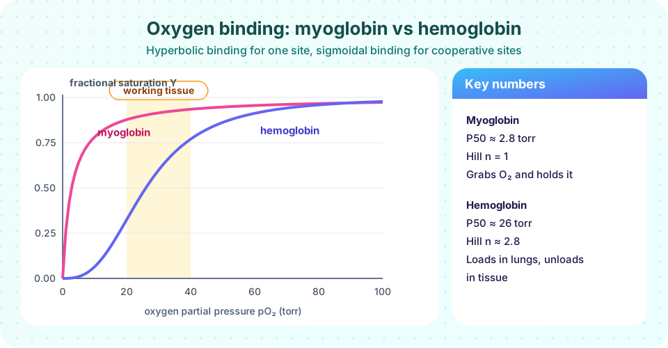
Myoglobin is a single polypeptide of about 153 residues with one heme group (an iron(II) held in a porphyrin ring). It is found in muscle and stores oxygen. Its P50 is about 2.8 torr (a few tenths of a kilopascal), so it is nearly saturated even when oxygen is scarce. Its binding curve is a simple hyperbola.
Hemoglobin: load and unload
Hemoglobin, in red blood cells, is a tetramer (two α and two β chains, each with a heme), so it can bind four O₂ molecules. Its P50 is about 26 torr and its curve is sigmoidal (S-shaped). The S-shape is the signature of positive cooperativity: the first oxygen binds with low affinity, but each additional one binds more easily. Max Perutz showed with X-ray crystallography that oxygen binding triggers a shift from the T (tense) deoxy form to the R (relaxed) oxy form. The structural change at one subunit is transmitted to the others through the subunit interfaces.
Why does this matter? Look at the pressures in the body (typical values): about 100 torr in the lungs, about 40 torr in resting tissue and as low as 20 torr in working muscle.
| Protein | Saturation at 100 torr | At 40 torr | At 20 torr | Oxygen released (100 → 40 torr) |
|---|---|---|---|---|
| Myoglobin | 0.97 | 0.93 | 0.88 | about 0.04 of capacity |
| Hemoglobin | 0.98 | 0.77 | 0.32 | about 0.21 of capacity |
(Values computed from the equations below with P50 = 2.8 and 26 torr and n = 1 and 2.8.) Myoglobin would hardly release any oxygen in the tissues; hemoglobin releases several times more, and in working muscle at 20 torr it delivers most of its load. Two proteins, one ligand, two jobs.
The Hill equation
A simple empirical description of cooperative binding is
Y = pO₂ⁿ / (P50ⁿ + pO₂ⁿ)
where n is the Hill coefficient. If n = 1 there is no cooperativity (a hyperbola); n > 1 gives positive cooperativity; n < 1 negative cooperativity. For hemoglobin n is about 2.8 to 3, below the theoretical maximum of 4 (the number of sites); it measures how cooperative the binding is, not the number of sites. Two classical models explain it structurally: the concerted (MWC) model, in which all subunits switch together between T and R, and the sequential (KNF) model, in which they switch one at a time. Real hemoglobin shows features of both.
Allosteric regulation of hemoglobin
Hemoglobin's affinity is tuned by effectors that bind elsewhere than the heme:
- H⁺ and CO₂ (the Bohr effect): in active tissue, lower pH and higher CO₂ stabilize the T state, shifting the curve right so more oxygen is released where it is needed.
- 2,3-bisphosphoglycerate (BPG): binds in the central cavity of the T state and lowers oxygen affinity. People living at high altitude have higher BPG levels. Fetal hemoglobin binds BPG less strongly, so it has a higher affinity than the mother's hemoglobin and takes oxygen across the placenta.
Common misconceptions
"A high Hill coefficient means more binding sites." It reflects cooperativity. The maximum n equals the number of sites but real values are lower.
"Hemoglobin binds oxygen more tightly than myoglobin." It binds more weakly (higher P50), which is exactly what allows it to give oxygen up.
"Binding strength is the same as binding specificity." Kd measures affinity; specificity is the ratio of affinities for the right and wrong ligands.
Try it
The first example plots both curves and computes oxygen delivery from lung to tissue pressures with the Hill equation.
Press Run (or Ctrl + Enter) to see the result here.
Program input · what you'd type while the program runs
Type the answers before you press Run, one per line. Each time the program reads a line (with input()), it gets the next line from this box.
The second example is interactive. Change the P50 and the Hill coefficient and watch how much oxygen is delivered between 100 torr (lungs) and 40 torr (tissue). It is a model of the Hill equation, not of a measured sample.
Check your understanding
Quick check · 6 questions
of 6 answered You scored / 6 · perfect!
-
1. In simple one-site binding, what does Kd equal?
From theta = [L]/(Kd + [L]), theta = 0.5 when [L] = Kd. -
2. Which has the lower P50 and therefore the higher oxygen affinity?
Myoglobin P50 is about 2.8 torr versus about 26 torr for hemoglobin. -
3. What does a sigmoidal binding curve indicate?
An S-shaped curve means binding at one site raises the affinity of the others. -
4. The Bohr effect means that:
H+ and CO2 stabilize the T state, shifting the curve right so oxygen is released in active tissue. -
5. A protein with Hill coefficient n = 1 shows:
n = 1 reduces the Hill equation to a hyperbola, i.e. independent binding sites. -
6. Fetal hemoglobin has a higher oxygen affinity than adult hemoglobin mainly because it:
Weaker BPG binding leaves fetal hemoglobin in a higher-affinity state, helping it take oxygen from maternal blood.
Summary
- Simple binding is hyperbolic:
θ = [L]/(Kd + [L]), with θ = 0.5 at [L] = Kd. - Myoglobin (P50 ≈ 2.8 torr, n = 1) stores oxygen; hemoglobin (P50 ≈ 26 torr, n ≈ 2.8) delivers it.
- Cooperativity arises from the T ⇌ R conformational change and is described by the Hill equation.
- H⁺, CO₂ and BPG allosterically lower hemoglobin's affinity, matching delivery to need.
Next: Chapter 3 review
Track your progress
Create a free account to tick off lessons and pick up where you left off on any device.
of Biochemistry lessons completed