


















Lesson 37 of 43 · Signaling and Regulation
Allostery and regulation of enzymes
Understand allosteric regulation, cooperative oxygen binding in hemoglobin, the MWC model, feedback inhibition and covalent control by phosphorylation and zymogen activation.
An enzyme that runs at a fixed speed is useless in a cell that needs to speed up, slow down and shut off. Cells tune proteins in several ways: by binding small molecules at sites away from the active site (allostery), by chemically modifying the protein (phosphorylation), by cutting a precursor into its active form, and by changing how much of the protein is made. This lesson looks at the first two in detail, using hemoglobin as the model and then metabolic pathways as the application.
What you will learn
- Define allostery and cooperativity and read a sigmoidal binding curve.
- Compare myoglobin and hemoglobin and explain how effectors such as 2,3-BPG and H⁺ shift the curve.
- Describe the concerted (MWC) and sequential models.
- Explain feedback inhibition and the main forms of covalent regulation.
Key terms
Allosteric regulation: control of a protein by a molecule binding at a site other than the active site, changing its conformation and activity.
Cooperativity: binding of one ligand changes the affinity of other sites on the same protein.
P₅₀: the oxygen partial pressure at which half of the binding sites are occupied.
Feedback inhibition: the end product of a pathway inhibits an early enzyme.
Zymogen: an inactive precursor of an enzyme that is activated by cutting (trypsinogen to trypsin).
Myoglobin and hemoglobin
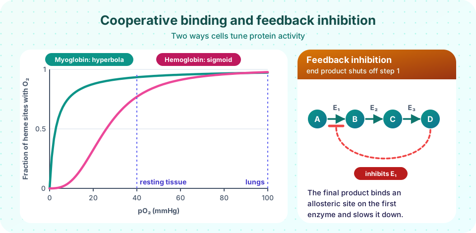
Myoglobin is a single-chain protein in muscle with one heme and so one oxygen-binding site. It binds oxygen along a hyperbolic curve, Y = pO₂ / (pO₂ + P₅₀), with a very low P₅₀ of about 2.8 mmHg: it holds oxygen tightly and stores it.
Hemoglobin, in red blood cells, is a tetramer (two α and two β chains) with four hemes. Its binding curve is sigmoidal, with P₅₀ about 26 mmHg and a Hill coefficient of about 2.8. The sigmoid shape is the signature of cooperativity: when one heme binds O₂, the protein changes shape and the other three bind more easily. The practical result is that hemoglobin is nearly saturated at lung pressures (about 100 mmHg) yet gives up a large fraction at the pressures found in working tissues. A protein with a hyperbolic curve and the same P₅₀ could not make such an efficient delivery.
The MWC (Monod–Wyman–Changeux) model explains the shape: the tetramer exists in two states, a low-affinity T (tense) state and a high-affinity R (relaxed) state, in equilibrium; oxygen binds R more tightly and so pulls the equilibrium toward R as more O₂ binds, and all subunits switch together. The sequential (Koshland–Némethy–Filmer) model instead lets subunits change one at a time. Real hemoglobin behaves like a mixture of both ideas.
Effectors shift the curve
- 2,3-Bisphosphoglycerate (2,3-BPG), made in red cells, binds in the central cavity of the T state and lowers oxygen affinity, shifting the curve right so more O₂ is released. Fetal hemoglobin (α₂γ₂) binds 2,3-BPG more weakly, so its affinity is higher and it can take oxygen from the mother's blood.
- H⁺ and CO₂ (the Bohr effect) also stabilize T: in active tissue, with more CO₂ and lower pH, hemoglobin releases more oxygen.
- Sickle cell hemoglobin has one amino acid change (Glu to Val, as in the mutation of the previous chapter) that makes deoxygenated molecules stick together into fibers.
Allosteric enzymes and feedback inhibition
Allosteric enzymes behave like hemoglobin: regulatory molecules bind a site away from the active site and shift the enzyme between active and inactive forms. Aspartate transcarbamoylase (ATCase), which starts pyrimidine synthesis, is inhibited by the end product CTP and activated by ATP. This is feedback inhibition: when the end product is plentiful it slows the first committed step, so the cell does not waste resources. PFK-1 in glycolysis is an allosteric enzyme controlled by ATP, AMP and citrate. Substrates for such enzymes often give sigmoidal rather than hyperbolic kinetics.
Covalent regulation
- Phosphorylation by kinases and dephosphorylation by phosphatases switch enzymes on or off within seconds. Glycogen phosphorylase is the classic case: phosphorylation converts the less active form b to the more active form a, tying it to hormone signaling (the previous lesson).
- Zymogen activation is irreversible: digestive proteases are made as inactive precursors and cut only where they are needed.
- Other modifications (acetylation, methylation, ubiquitin tagging) add further layers, and cells also control enzyme levels through gene expression and protein degradation.
These layers work on different time scales: allostery in milliseconds, phosphorylation in seconds to minutes, gene expression in minutes to hours.
Common misconceptions
"Allosteric means the molecule binds somewhere else, so it must be an inhibitor." Allosteric effectors can inhibit or activate.
"Cooperativity means the sites are close to each other." They are often far apart; the communication runs through conformational changes of the whole protein.
"Myoglobin is just a smaller hemoglobin." Their curves differ for a reason: myoglobin is built to store oxygen, hemoglobin to transport and release it.
Try it
Compare the two curves and the oxygen each can deliver between lungs (about 100 mmHg) and resting tissue (about 40 mmHg). The Hill equation is a model, so the numbers are estimates.
Press Run (or Ctrl + Enter) to see the result here.
Program input · what you'd type while the program runs
Type the answers before you press Run, one per line. Each time the program reads a line (with input()), it gets the next line from this box.
A toy MWC model for a four-site protein. The parameters are illustrative; they are chosen to show how the T/R equilibrium constant L controls cooperativity, not to fit real hemoglobin data.
Press Run (or Ctrl + Enter) to see the result here.
Program input · what you'd type while the program runs
Type the answers before you press Run, one per line. Each time the program reads a line (with input()), it gets the next line from this box.
Check your understanding
Quick check · 5 questions
of 5 answered You scored / 5 · perfect!
-
1. What is the shape of the oxygen-binding curve of hemoglobin?
Cooperative binding gives a sigmoidal curve; myoglobin's curve is hyperbolic. -
2. How does 2,3-BPG affect hemoglobin?
2,3-BPG stabilizes the T state and promotes oxygen release in tissues. -
3. In feedback inhibition, the inhibitor is usually
The end product shuts off an early enzyme so the pathway does not overproduce. -
4. Which type of regulation is irreversible?
Cutting peptide bonds cannot be undone, unlike phosphorylation or ligand binding. -
5. In the MWC model, a larger L (the T/R ratio without ligand) causes
When the T state dominates, the first ligand must pay a larger price to shift to R, giving a steeper sigmoid.
Summary
- Allosteric proteins are controlled by effectors binding away from the active site.
- Hemoglobin shows cooperativity (sigmoidal curve, P₅₀ about 26 mmHg); myoglobin is hyperbolic (P₅₀ about 2.8 mmHg).
- The MWC model describes T and R states; 2,3-BPG, H⁺ and CO₂ shift the curve to the right.
- Feedback inhibition, phosphorylation and zymogen activation add control on different time scales.
Next: Chapter 8 review
Track your progress
Create a free account to tick off lessons and pick up where you left off on any device.
of Biochemistry lessons completed