


















Lesson 12 of 43 · Amino Acids and Proteins
The peptide bond and the four levels of protein structure
Understand the planar peptide bond, backbone angles φ and ψ, the α-helix and β-sheet, and how primary, secondary, tertiary and quaternary structure build a protein.
Join amino acids end to end and you get a chain with a repeating backbone and a variable series of side chains. The backbone looks flexible, yet proteins fold into precise shapes. The reason lies in the peptide bond, which is rigid and flat, and in a few simple patterns of hydrogen bonding that the backbone can adopt: the α-helix and the β-sheet.
What you will learn
- Describe how a peptide bond forms and why the peptide group is planar.
- Interpret the backbone angles φ and ψ and the Ramachandran plot.
- Describe α-helices and β-sheets, including their hydrogen bonding.
- Define primary, secondary, tertiary and quaternary structure and the forces involved.
Key terms
Peptide bond: the amide bond between the carboxyl carbon of one amino acid and the amino nitrogen of the next, formed by condensation.
Polypeptide: a chain of amino acid residues, written from the N-terminus (free amino end) to the C-terminus (free carboxyl end).
φ and ψ: the rotation angles about the N–Cα bond (φ) and the Cα–C bond (ψ).
Secondary structure: regular local folding of the backbone, such as α-helices and β-sheets.
Domain: a compact, independently folding region of a protein, often 50 to 250 residues.
Forming the peptide bond
When the carboxyl carbon of one amino acid reacts with the amino nitrogen of another, water is released and a peptide bond forms. Every peptide has direction: the sequence is written from N-terminus to C-terminus, so Ala-Gly differs from Gly-Ala. The repeating backbone unit is N–Cα–C, and the side chains stick out from the α-carbons.
Why the peptide group is planar
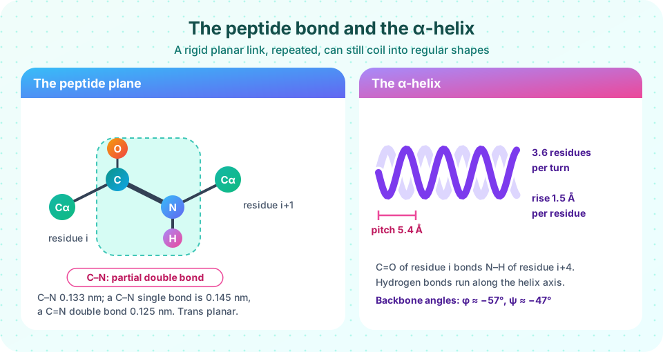
The C–N bond in a peptide has a length of about 0.133 nm, shorter than a typical C–N single bond (0.145 nm) and longer than a C=N double bond (0.125 nm). The reason is resonance: the nitrogen's lone pair is delocalized into the carbonyl group, giving the C–N bond partial double-bond character. A double bond cannot rotate freely, so the six atoms Cα, C, O, N, H and the next Cα lie in a rigid plane.
Two consequences follow:
- The peptide group is almost always trans (the two α-carbons on opposite sides of the C–N bond), because cis would bring the neighboring side chains too close together. The exception is the bond just before proline, where cis occurs in a small fraction of cases.
- The only real freedom in the backbone is rotation around the two bonds on either side of each plane.
φ, ψ and the Ramachandran plot
Rotation about N–Cα is φ (phi) and about Cα–C is ψ (psi). Most (φ, ψ) pairs would make atoms collide, so the allowed combinations are limited. G. N. Ramachandran plotted the allowed pairs in 1963 using hard-sphere models; the resulting diagram, the Ramachandran plot, shows that the allowed regions cluster in just a few areas:
- The right-handed α-helix: φ ≈ −57°, ψ ≈ −47°.
- The β-sheet region: φ ≈ −120°, ψ ≈ +130° (varies with strand type).
- A smaller region for the left-handed helix, which glycine, having no side chain, can populate more easily.
Glycine has the broadest allowed region; proline, whose ring locks φ near −63°, the narrowest.
The α-helix
In an α-helix the backbone coils like a spring. Key numbers:
- 3.6 residues per turn.
- A rise of 0.15 nm (1.5 Å) per residue, so 0.54 nm (5.4 Å) per turn.
- Hydrogen bonds link the C=O of residue i with the N–H of residue i + 4, running parallel to the helix axis.
- Side chains point outward from the helix, so every residue's backbone hydrogen-bonding groups are satisfied inside the helix, which is why helices are stable even in a nonpolar membrane.
The helix is right-handed in all natural proteins built from L-amino acids. Proline disrupts helices (its ring leaves no N–H to donate a hydrogen bond and restricts φ), and glycine's flexibility also makes it a poor helix former. A helix of 20 residues is about 3 nm long, enough to span a lipid bilayer.
The β-sheet
In a β-strand the backbone is nearly fully extended (about 0.33 to 0.35 nm per residue). Strands pair side by side, linked by hydrogen bonds between the backbone C=O of one strand and the N–H of its neighbor. In antiparallel sheets the strands run in opposite directions and the hydrogen bonds are nearly straight and strong; in parallel sheets they run in the same direction, with slightly angled bonds. The side chains alternate above and below the sheet, which gives many sheets a hydrophobic face and a polar face. Sheets are often slightly twisted.
Short turns (β-turns) that reverse the direction of the chain connect helices and strands.
The four levels of structure
| Level | What it describes | Main forces |
|---|---|---|
| Primary | the amino acid sequence | covalent peptide bonds (and disulfide bonds) |
| Secondary | local helices, sheets, turns | backbone hydrogen bonds |
| Tertiary | the full 3D fold of one chain | hydrophobic effect, H-bonds, salt bridges, van der Waals, disulfides, metal binding |
| Quaternary | assembly of several chains (subunits) | same noncovalent forces |
A protein such as hemoglobin has quaternary structure: two α chains and two β chains, each folded around a heme group. Many enzymes are dimers or larger oligomers. The overall shape divides proteins into globular types (compact, soluble: enzymes, antibodies) and fibrous types (long and rod-like: collagen's triple helix, keratin's coiled coils, silk's β-sheets).
Common misconceptions
"The peptide bond rotates freely." It has partial double-bond character and is planar and mostly trans. The rotations occur at φ and ψ.
"Hydrogen bonds in helices are between side chains." They are between backbone C=O and N–H groups; side chains point outward.
"Secondary structure comes after tertiary structure." The levels are descriptive categories, not a timeline. Local structure and global fold form together during folding.
Try it
First, a schematic Ramachandran plot. The shaded regions are approximate; real plots are drawn from thousands of experimental structures.
Press Run (or Ctrl + Enter) to see the result here.
Program input · what you'd type while the program runs
Type the answers before you press Run, one per line. Each time the program reads a line (with input()), it gets the next line from this box.
Second, a helical wheel. Residues in an α-helix are spaced 100° apart around the axis (360° ÷ 3.6). Viewing a sequence from the end shows whether one side of the helix is hydrophobic and the other polar, a pattern called amphipathic. This is a demonstration of the geometry.
Check your understanding
Quick check · 6 questions
of 6 answered You scored / 6 · perfect!
-
1. Why is the peptide bond planar?
Delocalization of the nitrogen lone pair into the carbonyl gives the C-N bond partial double-bond character, preventing rotation. -
2. How many residues are there per turn of an alpha helix?
3.6 residues per turn; 5.4 is the pitch in angstroms. -
3. In an alpha helix, the C=O of residue i hydrogen-bonds to the N-H of residue:
The helix is stabilized by i to i+4 hydrogen bonds along the axis. -
4. Which amino acid is a strong helix breaker because it has no N-H and a rigid ring?
Proline cannot donate a backbone hydrogen bond and restricts phi, so it kinks helices. -
5. Hemoglobin has two alpha and two beta chains. That is an example of:
Several polypeptide subunits assembling into one complex is quaternary structure. -
6. In which structure do side chains point alternately above and below the plane?
In a beta strand, consecutive side chains alternate sides of the sheet.
Summary
- The peptide bond is planar and mostly trans because of partial double-bond character; backbone rotation happens at φ and ψ.
- The Ramachandran plot shows the allowed (φ, ψ) combinations; helices and sheets occupy distinct regions.
- α-helix: 3.6 residues per turn, 5.4 Å pitch, i → i+4 hydrogen bonds. β-sheet: extended strands joined by interstrand hydrogen bonds.
- Primary to quaternary structure builds a protein; the hydrophobic effect and weak forces hold tertiary and quaternary structure.
Track your progress
Create a free account to tick off lessons and pick up where you left off on any device.
of Biochemistry lessons completed