


















Lesson 22 of 45 · Sensory Systems
Vision: from photons to V1
Follow light through the eye, the rods and cones, center-surround ganglion cells, the optic chiasm, the LGN and primary visual cortex, with simple models.
Vision is the sense we know best, and it takes up a large share of the cortex. The path from the world to your visual experience has clear stages: optics, a thin sheet of neural tissue (the retina), a relay in the thalamus and a series of cortical areas. At each stage the code becomes more abstract: from light intensity, to contrast, to edges, to objects.
What you will learn
- How the eye focuses light and how photoreceptors transduce it
- Why ganglion cells report contrast and not brightness
- How signals reach and are organized in the primary visual cortex
- What the dorsal and ventral streams do
Key terms
Photoreceptor: a rod or cone, the retinal cell that absorbs light.
Fovea: the central pit of the retina, packed with cones, giving the sharpest vision.
Receptive field: the part of the visual field that changes a neuron's firing.
LGN: the lateral geniculate nucleus, the thalamic relay for vision.
V1: primary visual cortex, in the occipital lobe.
Optics: getting the image onto the retina
Light is refracted mainly by the cornea, which provides about two thirds of the eye's focusing power. The lens adds adjustable power: ciliary muscles change its shape so you can focus near or far (accommodation). The pupil controls how much light gets in. The image lands upside down on the retina, and nothing needs to "flip it back": the brain simply learns the mapping.
The retina: a piece of brain in the eye
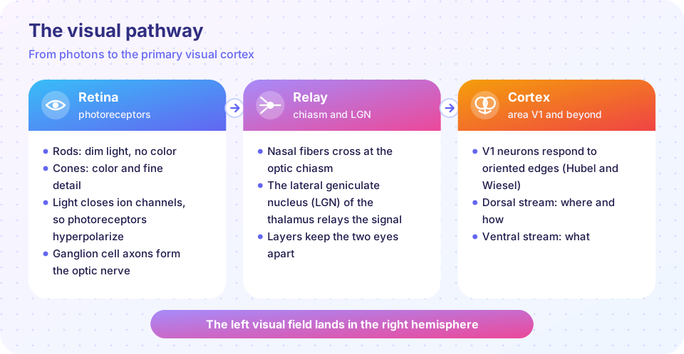
The retina is a layered sheet of neurons. Light passes through most of the layers before reaching the photoreceptors at the back. The signal then goes forward: photoreceptor, bipolar cell, ganglion cell. Horizontal and amacrine cells connect sideways and sharpen the signal. Ganglion cell axons bundle into the optic nerve.
There are about 120 million rods and about 6 million cones in each human eye, in round numbers. Rods are extremely sensitive and work in dim light, but give no color. Cones need more light and give color and fine detail. The fovea contains cones almost exclusively, which is why you must look directly at a small word to read it.
Phototransduction
The photoreceptor story is surprising: light makes the cell hyperpolarize, not depolarize. In the dark, cGMP holds sodium-permitting channels open (the "dark current"), so the cell sits near -40 mV and releases glutamate continuously. A photon isomerizes the pigment (rhodopsin in rods). The pigment activates a G-protein (transducin), which activates an enzyme (phosphodiesterase) that breaks down cGMP. Channels close, the cell hyperpolarizes, and glutamate release falls. A single photon can produce a measurable response in a rod, and this multi-step amplification is why.
Color
Humans have three cone types with peak sensitivity near 420 nm (S, short), about 530 nm (M, medium) and about 560 nm (L, long). Color is read from the ratios of the three responses. This is the trichromatic theory of Young and Helmholtz. Downstream cells compare cones in opponent pairs (red versus green, blue versus yellow), which explains why there is no "reddish green" in experience. Color-vision deficiencies usually come from missing or shifted L or M pigments, which is why they are more common in males: the genes sit on the X chromosome.
Ganglion cells: reporting contrast
Stephen Kuffler recorded from cat retinal ganglion cells in 1953 and found center-surround receptive fields. An on-center cell fires most when light hits a small central spot and the surrounding ring is dark. Light in the ring inhibits it. A uniform patch of light covering both gives only a weak response. The cell therefore reports differences in light, i.e. edges and contrast, and not the absolute amount. This compresses the signal: about 6 million cones feed only about one million ganglion cells.
A useful model is the difference of two Gaussians, a narrow excitatory center minus a broader inhibitory surround.
Relay and cortex
At the optic chiasm, fibers from the nasal half of each retina cross. After the crossing, the right optic tract carries information about the left visual field from both eyes. It ends in the lateral geniculate nucleus (LGN) of the thalamus, whose layers keep inputs from the two eyes separate. LGN neurons look much like ganglion cells (center-surround), and they project through the optic radiations to V1.
In 1959 to 1962, David Hubel and Torsten Wiesel recorded from cat V1 and found neurons that fire for a bar of light at a particular orientation. Some are simple cells (a bar must fall at a precise position), others complex cells (the bar can shift). V1 is also organized into columns of cells sharing the same orientation preference and eye preference. They shared the 1981 Nobel Prize for this work. V1 is a map: the fovea is greatly magnified, taking up a disproportionate share of the cortical area.
Beyond V1, two broad streams emerge. The ventral stream (toward the temporal lobe) identifies what something is, including faces and objects. The dorsal stream (toward the parietal lobe) handles where it is and how to act on it. Goodale and Milner's studies of a patient with ventral damage, who could not name the orientation of a slot but could post a letter through it, support this "perception versus action" view.
Common misconceptions
"Rods and cones are depolarized by light." They hyperpolarize. The neurotransmitter signal falls when light arrives.
"We see in the eye." Much of the "seeing" is cortical. People with intact eyes and V1 damage can report blindness in part of the visual field, while some respond to stimuli there without awareness ("blindsight").
"The brain flips the upside-down image." There is no flip step: the brain learns to interpret the map.
Try it
Press Run (or Ctrl + Enter) to see the result here.
Program input · what you'd type while the program runs
Type the answers before you press Run, one per line. Each time the program reads a line (with input()), it gets the next line from this box.
Check your understanding
Quick check · 5 questions
of 5 answered You scored / 5 · perfect!
-
1. What happens to a photoreceptor when it absorbs light?
Light closes cGMP-gated channels, so the cell hyperpolarizes and glutamate release falls. -
2. Why can a ganglion cell with a center-surround field ignore uniform light?
Opposing center and surround means the cell signals contrast and edges. -
3. Which pathway carries information about the left visual field from both eyes?
After the chiasm, the right optic tract carries the left visual field to the right hemisphere. -
4. What did Hubel and Wiesel discover in cat V1?
Simple and complex cells respond to bars of a preferred orientation. -
5. The ventral stream mostly answers which question?
The ventral (temporal) stream supports object and face identification.
Summary
- The cornea and lens focus light on the retina, where rods and cones transduce it.
- Photoreceptors hyperpolarize in light; three cone types give color through ratios.
- Ganglion cells have center-surround fields and report contrast.
- Signals cross at the chiasm, relay in the LGN and reach V1, where cells prefer oriented edges.
- Ventral and dorsal streams handle "what" and "where or how".
Next: Hearing and balance
Track your progress
Create a free account to tick off lessons and pick up where you left off on any device.
of Neuroscience lessons completed