


















Lesson 27 of 45 · Motor Systems
Spinal cord and reflexes
Learn how the spinal cord acts alone: the knee-jerk reflex, muscle spindles, Golgi tendon organs, withdrawal reflexes and central pattern generators for walking.
Touch a hot pan and your hand is gone before you feel the burn. That speed comes from circuits inside the spinal cord, which can produce useful movements without waiting for a decision from the brain. This lesson explains the best-known reflex, the sensors behind it, and the spinal circuits that generate rhythms such as walking.
What you will learn
- How the knee-jerk reflex works, step by step
- What muscle spindles and Golgi tendon organs sense
- How reciprocal inhibition and withdrawal reflexes coordinate muscles
- What central pattern generators are
Key terms
Muscle spindle: a stretch sensor inside a muscle.
Golgi tendon organ: a force sensor in the tendon.
Monosynaptic: with only one synapse between sensory and motor neuron.
Reciprocal inhibition: excitation of one muscle with inhibition of its opponent.
Central pattern generator (CPG): a spinal circuit that produces rhythmic output without rhythmic input.
The stretch reflex
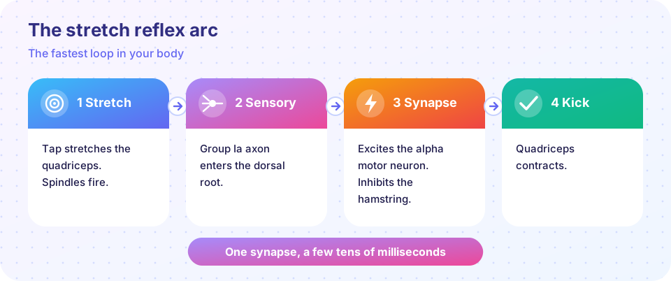
A reflex doctor taps the patellar tendon just below the kneecap. This briefly stretches the quadriceps. Inside the muscle, muscle spindles (bundles of specialized intrafusal fibers) notice the stretch. Their group Ia sensory axons enter the spinal cord through the dorsal root and synapse directly onto the alpha motor neurons of the same muscle. The motor neurons fire, the quadriceps contracts, and the leg kicks. Because only one synapse lies between sensor and effector, it is called monosynaptic and the delay is only a few tens of milliseconds.
At the same time, branches of the Ia axons excite an inhibitory interneuron that silences the motor neurons of the opposing hamstring. This is reciprocal inhibition, so the antagonist does not fight the movement.
Why have the reflex at all? It stabilizes posture. If a load suddenly stretches a muscle, the reflex immediately increases the muscle's force. Doctors use it clinically: exaggerated reflexes, or none at all, help localize damage to the pathway or to the upper motor neurons. (This is educational background, not a way to diagnose yourself.)
Gamma motor neurons
The spindle must keep working when the muscle shortens. Gamma motor neurons contract the ends of the intrafusal fibers so the spindle remains taut and sensitive. The cord therefore co-activates alpha and gamma motor neurons during voluntary movement.
The opposite sensor: Golgi tendon organs
Golgi tendon organs sit at the junction of muscle and tendon and sense force through group Ib axons. Via interneurons they inhibit the muscle's own motor neurons. They were once thought to be a mere emergency brake against overload; today they are considered part of a continuous force-feedback system that helps smooth movements.
Withdrawal and crossed extension
If you step on a sharp object, nociceptive afferents excite spinal interneurons that activate flexor muscles of that leg (the flexion withdrawal reflex) and, on the opposite side, extensors to support your weight (crossed extension). It takes more than one synapse (it is polysynaptic), is slower than the knee jerk, and the pain you feel arrives after you already moved.
Rhythms in the cord
Thomas Graham Brown showed in 1911 that the cat spinal cord, cut off from the brain, could produce alternating flexor-extensor bursts. He proposed a half-center model: two groups of neurons inhibit each other and alternate. We now call such circuits central pattern generators. They produce the basic timing of walking and swimming; sensory feedback adjusts it for the ground, and descending commands start, stop and steer. Humans have comparable spinal circuits, which is one reason rehabilitation after spinal cord injury uses repetitive stepping practice.
Spinal cord injury in brief
The level of an injury determines what is lost. Damage cuts descending commands (weakness or paralysis below the level) and ascending sensation (numbness below the level). Right after a severe injury the cord goes through spinal shock with absent reflexes; later reflexes return, often exaggerated, because descending inhibition is missing.
Common misconceptions
"Reflexes bypass the brain entirely." The brain is informed and can modulate reflexes: you can brace to reduce a tap's effect, and training can change reflex size.
"The knee-jerk reflex tests muscle strength." It tests the sensory-motor loop and its descending control, not strength.
Try it
Press Run (or Ctrl + Enter) to see the result here.
Program input · what you'd type while the program runs
Type the answers before you press Run, one per line. Each time the program reads a line (with input()), it gets the next line from this box.
Check your understanding
Quick check · 5 questions
of 5 answered You scored / 5 · perfect!
-
1. Why is the knee-jerk reflex called monosynaptic?
The Ia axon synapses directly on the alpha motor neuron. -
2. What do Golgi tendon organs mainly sense?
They report tension via group Ib axons. -
3. What does reciprocal inhibition do in the knee jerk?
Inhibitory interneurons reduce antagonist activity so the movement is not opposed. -
4. What is a central pattern generator?
CPGs create the alternating bursts used in walking and swimming. -
5. Why does gamma motor neuron activity matter?
Contracting spindle ends keeps the stretch sensor taut.
Summary
- The stretch reflex is a monosynaptic loop from spindle to alpha motor neuron.
- Reciprocal inhibition coordinates agonist and antagonist.
- Golgi tendon organs report force; gamma motor neurons keep spindles sensitive.
- Withdrawal reflexes are polysynaptic; central pattern generators drive rhythms.
Track your progress
Create a free account to tick off lessons and pick up where you left off on any device.
of Neuroscience lessons completed