


















Lesson 28 of 45 · Motor Systems
Cortex, basal ganglia and cerebellum
See how the motor cortex commands movement, how basal ganglia choose actions through go and stop pathways, and how the cerebellum corrects errors.
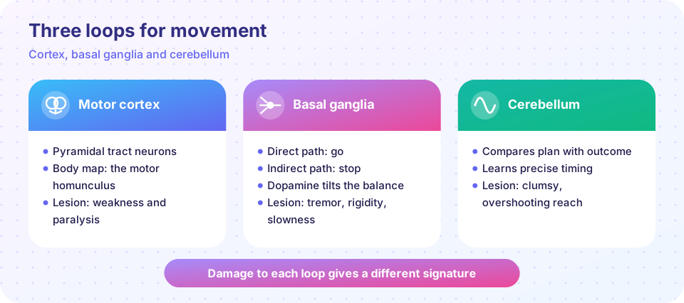
Reflexes are simple. Playing tennis is not. Voluntary movement needs a plan, a choice among competing actions, precise timing and constant correction. Three systems share the job: the motor cortex, the basal ganglia and the cerebellum. Each gives a recognizable signature when it is damaged, which is how neurologists learned what they do.
What you will learn
- How the motor cortex represents movement and sends commands
- How the direct and indirect basal ganglia pathways select actions
- How the cerebellum learns to correct and time movements
- What damage to each structure looks like
Key terms
Corticospinal tract: the main descending pathway from motor cortex to the spinal cord.
Striatum: the input stage of the basal ganglia (caudate and putamen).
Substantia nigra pars compacta: dopamine neurons projecting to the striatum.
Purkinje cell: the large cerebellar output neuron of the cortex of the cerebellum.
Forward model: a prediction of the sensory consequences of a motor command.
The motor cortex
Primary motor cortex (M1) lies in the precentral gyrus. In the 1930s to 1950s, Wilder Penfield stimulated cortex in patients and mapped a motor homunculus: neighboring cortex controls neighboring body parts, with large areas for the hands and face. Pyramidal neurons in layer V send axons down the corticospinal tract. About 90 percent of the fibers cross (decussate) in the lower medulla, so the right motor cortex controls the left side of the body.
Movement direction is not encoded by single cells. In the 1980s, Apostolos Georgopoulos recorded from monkey M1 and found that each cell fires most for a preferred direction but broadly; the population vector, the weighted sum across cells, accurately predicts the reach direction. This idea is the basis of many brain-computer interfaces. Planning areas lie in front of M1 (premotor and supplementary motor cortex) and in the parietal lobe. Some premotor neurons in macaques (area F5, studied by Giacomo Rizzolatti's group) fire both when the monkey performs a grasp and when it watches one. These mirror neurons are a lively topic; how far they explain human understanding of actions is debated.
The basal ganglia: choosing and starting
The basal ganglia include the striatum, the globus pallidus (external, GPe, and internal, GPi), the subthalamic nucleus (STN) and the substantia nigra. They receive input from the whole cortex and send output through the thalamus back to the cortex. Their rule of thumb: GPi neurons fire tonically and brake the thalamus. Two pathways change that brake:
- Direct pathway ("go"): striatal neurons with D1 dopamine receptors inhibit GPi. This releases the brake on the thalamus, facilitating a movement.
- Indirect pathway ("stop"): striatal neurons with D2 receptors inhibit GPe, which indirectly strengthens GPi's brake, suppressing competing movements.
Dopamine from the substantia nigra excites the direct pathway (D1) and inhibits the indirect pathway (D2), so more dopamine favors action. The model is a simplification (the pathways cooperate and are active together), but it explains why a loss of dopamine causes slow movement and why too much dopamine activity relates to excess movement. The basal ganglia also contribute to habit learning and to reward-based choice.
The cerebellum: correcting and timing
The cerebellum holds most of the brain's neurons (on the order of 80 percent) in about a tenth of its volume. Its cortex has a very regular design: mossy fibers drive granule cells, whose parallel fibers contact huge Purkinje cells, the only output of the cerebellar cortex. Climbing fibers from the inferior olive wrap around each Purkinje cell and signal errors. In the Marr, Albus and Ito theory, error signals from the climbing fibers weaken parallel-fiber synapses that were active when an error occurred, so the next movement is better.
The cerebellum acts like a machine that holds a forward model: it predicts the consequence of a command and compares it with feedback. Classic evidence is prism adaptation: a person throwing darts wearing prisms misses at first and then, over a few dozen throws, corrects; on removing the prisms they miss in the opposite direction. Patients with cerebellar damage adapt poorly.
Signatures of damage
| Region | Typical problem |
|---|---|
| Motor cortex or tract | weakness or paralysis on the opposite side |
| Basal ganglia (dopamine loss) | slowness, rigidity, rest tremor |
| Cerebellum | clumsy, overshooting reaching, unsteady wide-based walking |
Common misconceptions
"The cerebellum is just for balance." It also contributes to precise timing, learning of skills and aspects of cognition.
"Basal ganglia pathways are a simple on and off switch." Real circuits are richer; "go" and "stop" is a teaching model.
Try it
Press Run (or Ctrl + Enter) to see the result here.
Program input · what you'd type while the program runs
Type the answers before you press Run, one per line. Each time the program reads a line (with input()), it gets the next line from this box.
Press Run (or Ctrl + Enter) to see the console output here.
No output.
Check your understanding
Quick check · 5 questions
of 5 answered You scored / 5 · perfect!
-
1. Which tract carries the main command from motor cortex to the spinal cord?
Layer V pyramidal neurons form the corticospinal tract. -
2. What does dopamine do to the direct pathway?
D1 receptors on direct pathway neurons are excitatory. -
3. What do climbing fibers signal to Purkinje cells?
In the Marr-Albus-Ito theory, they provide teaching signals about errors. -
4. A patient reaches for a cup and overshoots, with an unsteady wide-based walk. Which region is most likely involved?
Overshoot and wide-based gait are classic cerebellar signs. -
5. Roughly how many corticospinal fibers cross in the medulla?
Most cross, so each hemisphere controls the opposite side of the body.
Summary
- Motor cortex encodes movements in populations of neurons and projects via the corticospinal tract.
- Basal ganglia use dopamine-modulated direct and indirect pathways to select actions.
- The cerebellum predicts, compares and learns, improving accuracy and timing.
- Each structure has a characteristic pattern of impairment.
Next: Chapter 6 review
Track your progress
Create a free account to tick off lessons and pick up where you left off on any device.
of Neuroscience lessons completed