


















Lesson 17 of 45 · Synapses and Neurotransmitters
The chemical synapse: release, receptors and cleanup
Follow neurotransmission step by step: calcium entry, SNARE-mediated vesicle fusion, quantal release, receptor binding and the removal of transmitter from the cleft.
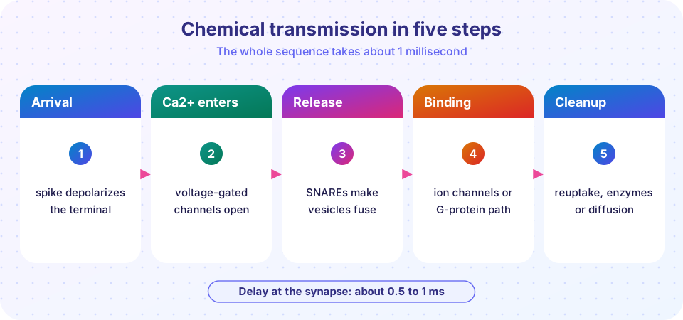
When a spike reaches the end of an axon, it triggers one of the fastest and most carefully controlled chemical events in biology. Within about half a millisecond, tiny packages of transmitter are released, cross a gap 20 nanometers wide and open channels on the next cell. The sequence is the same, with variations, at billions of synapses. Understanding each step shows you how drugs, toxins and diseases can interfere with the nervous system.
What you will learn
- The five steps from presynaptic spike to postsynaptic response.
- How calcium triggers vesicle fusion, and the role of SNARE proteins.
- What quantal release means, and how Bernard Katz discovered it.
- How transmitter is removed, and how toxins and drugs interfere.
Key terms
- Vesicle: a membrane bubble about 40 nm wide that stores transmitter.
- Active zone: the part of the presynaptic membrane where vesicles dock and release.
- SNARE proteins: proteins that pull the vesicle and plasma membranes together so they fuse.
- Quantum: the transmitter content of one vesicle, producing the smallest unit of response.
- Reuptake: transporter proteins pump transmitter back into cells.
Step 1: the spike arrives
The action potential depolarizes the presynaptic terminal. The terminal contains neurotransmitter packed into vesicles and a cluster of voltage-gated calcium channels next to the active zone.
Step 2: calcium enters
The depolarization opens voltage-gated Ca²⁺ channels. Calcium is about 10,000 times more concentrated outside the cell than inside (2 mM against 200 nM, from Chapter 3), so it floods into the terminal. Local calcium concentration near the channel mouth briefly rises to tens of micromolar, which is enough to trigger release. Calcium is the essential trigger: if you remove it from the bathing solution, a spike arrives at the terminal but nothing is released. Release depends steeply on calcium, roughly with the fourth power of the concentration in many synapses, which means that a small change in calcium entry changes release a lot.
Step 3: vesicles fuse and release
Docked vesicles are held in place by a set of SNARE proteins: synaptobrevin (on the vesicle) and syntaxin and SNAP-25 (on the plasma membrane) twist into a tight bundle, like a zipper that is waiting to be closed. A protein called synaptotagmin acts as the calcium sensor. When Ca²⁺ binds, it triggers the SNARE zipper to close and the vesicle membrane fuses with the plasma membrane, opening a pore through which the transmitter diffuses out. This takes a fraction of a millisecond.
This system is a target for natural toxins. Botulinum toxin cleaves SNARE proteins at the neuromuscular junction, so acetylcholine release fails and muscles are paralyzed. (Tiny, controlled doses are used medically for muscle spasms and in cosmetic treatments.) Tetanus toxin is carried back into the spinal cord and blocks release from inhibitory neurons, which leads to uncontrolled muscle contraction.
Quantal release: Katz's discovery
In the 1950s, Bernard Katz and his colleagues recorded from the frog neuromuscular junction at rest and saw tiny, equal-sized blips about 0.4 mV, which they called miniature end-plate potentials. When a nerve spike arrived, the response was a whole-number multiple of this unit. The conclusion: transmitter is released in packets, or quanta, which turned out to be the contents of single vesicles. A vesicle at the neuromuscular junction holds a few thousand acetylcholine molecules. Katz received the 1970 Nobel Prize for this and related work.
At most synapses, the average response to a spike is described by three numbers: the number of release sites (n), the probability that each releases (p), and the size of the response to one vesicle (q). The mean is n × p × q, and individual trials vary, including complete failures when no vesicle is released. Typical release probabilities of central synapses vary widely, from a few percent to over 50%.
Step 4: transmitter binds receptors
The transmitter diffuses across the 20 nm cleft in a fraction of a microsecond and binds receptors on the postsynaptic membrane. Two receptor classes exist (see the next lesson):
- Ionotropic receptors are ion channels themselves. Binding opens the channel within about a millisecond, so the effect is fast.
- Metabotropic receptors are G-protein-coupled receptors. They trigger a chain of intracellular reactions, so the effect is slower, longer and can reach well beyond the synapse.
Opening channels lets ions flow, changing the postsynaptic membrane potential. This change is called a postsynaptic potential.
Step 5: cleanup
If transmitter lingered, the signal would blur. Several mechanisms clear it:
- Reuptake: transporters carry transmitter back into the terminal or into astrocytes. Glutamate transporters in astrocytes are a major route; dopamine and serotonin transporters recycle those transmitters.
- Enzymatic breakdown: acetylcholinesterase in the cleft splits acetylcholine within a fraction of a millisecond. Monoamine oxidase and COMT degrade monoamines.
- Diffusion: the transmitter simply spreads away.
Many drugs work by interfering with these steps, such as drugs that slow reuptake of serotonin or inhibit acetylcholinesterase. This lesson gives information about mechanisms only; it is not medical advice.
Vesicle membrane is also recycled. After fusion, the membrane is retrieved by endocytosis, often using the protein clathrin, and the vesicle is refilled by transporters in its membrane, all within seconds to tens of seconds.
Synaptic delay
From spike arrival to the start of the postsynaptic response takes about 0.5 to 1 ms at body temperature. Most of this time goes to calcium channel opening and vesicle fusion. The delay adds up along multi-synapse paths, which is one reason fast reflexes use few synapses.
Common misconceptions
"Neurotransmitter release is continuous." It is mostly triggered by spikes, although spontaneous single-vesicle events (miniatures) also occur at low rates.
"Every spike releases transmitter." At many central synapses the release probability is below 50%, so each spike has a good chance of failing.
"The transmitter decides excitation or inhibition." The receptor on the target does. Acetylcholine excites skeletal muscle through nicotinic receptors but slows the heart through muscarinic receptors.
Try it
Press Run (or Ctrl + Enter) to see the result here.
Program input · what you'd type while the program runs
Type the answers before you press Run, one per line. Each time the program reads a line (with input()), it gets the next line from this box.
Check your understanding
Quick check · 5 questions
of 5 answered You scored / 5 · perfect!
-
1. Which ion entering the terminal triggers vesicle release?
Calcium entry through voltage-gated channels triggers fusion. -
2. What does botulinum toxin do?
By cleaving SNAREs it prevents vesicle fusion at the neuromuscular junction. -
3. A quantum of transmitter corresponds to...
Katz showed that release occurs in packets equal to single vesicles. -
4. Which receptor type produces the faster response?
Ionotropic receptors are channels and respond within about a millisecond. -
5. Acetylcholinesterase...
It is the enzyme that terminates cholinergic signals.
Summary
- A spike opens Ca²⁺ channels; Ca²⁺ binds synaptotagmin and SNAREs fuse vesicles, releasing transmitter in quanta.
- Transmitter binds ionotropic (fast) or metabotropic (slow) receptors.
- Reuptake, enzymes and diffusion end the signal.
- Toxins, drugs and diseases can interfere with any step.
Next: Neurotransmitters
Track your progress
Create a free account to tick off lessons and pick up where you left off on any device.
of Neuroscience lessons completed