


















Lesson 4 of 45 · Getting Started
Brain regions and how we study them
Tour the major brain regions, from cortex lobes to brainstem, then compare the methods scientists use to study them, from patch clamp to fMRI.
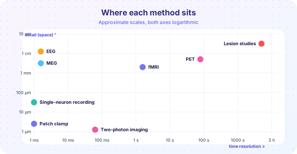
The brain is not a uniform lump. It is divided into regions with different structure, connections and jobs. This lesson gives you a guided tour of the major parts, then turns to a question that shapes all of neuroscience: how do we know what any region does? The tools used to answer that question all trade off detail in space against speed in time.
What you will learn
- The three main divisions of the brain and their main structures.
- What the four lobes of the cortex are mostly specialized for.
- The roles of the thalamus, hypothalamus, basal ganglia, hippocampus and amygdala.
- How the main research methods differ in space and time resolution.
Key terms
- Cerebral cortex: the folded outer layer of the cerebrum, about 2 to 4 mm thick.
- Gyrus / sulcus: a ridge or a groove of the folded cortex.
- Lesion: damage to a brain region, from injury, stroke or surgery.
- Spatial resolution: how small a region a method can distinguish.
- Temporal resolution: how quickly a method can detect a change.
The big divisions
Embryologists divide the brain into three parts that you can still recognize in adults.
- Forebrain: the cerebrum (two hemispheres of cortex plus deep structures) and the diencephalon (thalamus and hypothalamus).
- Midbrain: a small region handling eye movements, reflexes to sights and sounds, and dopamine pathways.
- Hindbrain: the pons, medulla and cerebellum.
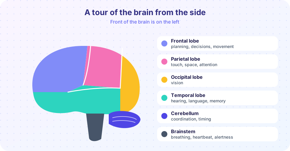
The brainstem is the midbrain, pons and medulla together. It controls breathing, heart rate, sleep and alertness, so brainstem damage is far more dangerous than damage to most cortical areas.
The cerebral cortex and its lobes
The cortex is where a great deal of perception, language, planning and awareness happens. Each hemisphere has four lobes.
- Frontal: movement planning and execution (motor cortex), decisions, working memory, speech production (Broca's area, usually on the left).
- Parietal: touch, body position and spatial attention.
- Temporal: hearing, recognizing objects, language comprehension (Wernicke's area, usually on the left) and memory.
- Occipital: the first stages of vision.
The cortex is folded so that a large sheet fits in a skull. Spread out, it covers roughly 2,000 cm² (the figure varies between people). Most of it is neocortex, arranged in six layers of neurons and organized in vertical columns that process related information. The hemispheres are joined by the corpus callosum, a bundle of about 200 million axons.
Deep structures
Under the cortex sit structures you will meet repeatedly.
| Structure | Main roles |
|---|---|
| Thalamus | Relay station: almost all sensory information (except smell) passes through it on the way to cortex |
| Hypothalamus | Hunger, thirst, temperature, sleep cycles and hormone control through the pituitary |
| Basal ganglia | Selecting and starting movements; affected in Parkinson's disease |
| Hippocampus | Forming new memories of facts and events; spatial navigation |
| Amygdala | Emotional learning, especially fear and threat detection |
| Cerebellum | Fine-tuning movement, timing and balance; holds about 80% of all neurons |
Remember the warning from the last lesson: no region works alone. "The amygdala is the fear center" is a handy slogan, but fear also needs sensory cortex, hippocampus, the hypothalamus and the brainstem.
How do we know what a region does?
Neuroscientists combine several kinds of evidence.
- Lesion and neuropsychology. If damage to a region removes an ability, the region is probably needed for it. Broca's and H.M.'s cases are classics. The limit: damage is rarely neat, and other regions may compensate.
- Recording electrical activity. A tiny electrode can record from a single neuron (spikes), a glass pipette can clamp one cell's membrane (patch clamp), and electrodes on the scalp record the summed activity of millions of neurons (EEG). A magnetic version of EEG is MEG.
- Imaging. MRI shows structure in fine detail. fMRI measures the BOLD signal, a change in blood oxygen that follows neural activity by a few seconds. PET follows radioactive tracers to map metabolism or receptors.
- Stimulation. Transcranial magnetic stimulation (TMS) uses a magnetic pulse to briefly disturb a cortical area in a person.
- Optogenetics and imaging in animals. Light-sensitive proteins let scientists switch specific cell types on and off with light (developed in the mid-2000s by teams including Karl Deisseroth and Ed Boyden), and two-photon microscopy films activity in individual cells in living animals.
Each method answers a different question. Recording tells you what correlates with a task. Lesions and stimulation tell you what is necessary or sufficient. Using several together is how the field avoids being fooled by any one.
Common misconceptions
"fMRI shows neurons firing." It shows blood oxygenation changes, an indirect and slow signal. Bright spots mean more oxygenated blood flow to that area, and the link to spiking is real but loose.
"If a region lights up, it is the region for that task." Many areas respond to many tasks. Correlation in imaging is not proof of necessity.
"The brain's folds are decoration." Folding increases cortical surface area, which is tied to computing capacity.
Try it
Press Run (or Ctrl + Enter) to see the result here.
Program input · what you'd type while the program runs
Type the answers before you press Run, one per line. Each time the program reads a line (with input()), it gets the next line from this box.
Check your understanding
Quick check · 5 questions
of 5 answered You scored / 5 · perfect!
-
1. Which structure relays almost all sensory information to the cortex?
The thalamus is the main relay. Smell is the exception, reaching cortex without a thalamic relay. -
2. Which lobe contains primary visual cortex?
The occipital lobe at the back of the brain receives the first stages of vision. -
3. What does the BOLD signal in fMRI measure?
BOLD follows blood oxygen changes linked to neural activity, a slower and indirect signal. -
4. Which method has fast timing but poor spatial detail?
EEG has millisecond timing but a spatial resolution of centimeters at best. -
5. Where do most of the brain's neurons live?
The cerebellum holds about four in five neurons.
Summary
- The brain divides into forebrain, midbrain and hindbrain; the brainstem handles life-support functions.
- The cortex has frontal, parietal, temporal and occipital lobes with rough specialties.
- Thalamus, hypothalamus, basal ganglia, hippocampus, amygdala and cerebellum are the key deep structures.
- Methods trade space against time: patch clamp and microscopy are detailed, EEG and MEG are fast, fMRI and PET are slower.
- Lesion, recording and stimulation studies answer different questions, so strong conclusions use several.
Next: Chapter 1 review
Track your progress
Create a free account to tick off lessons and pick up where you left off on any device.
of Neuroscience lessons completed