


















Lesson 23 of 45 · Sensory Systems
Hearing and balance
Trace sound from the eardrum to the cochlea, hair cell transduction and sound localization, then see how the vestibular system senses head motion.
Sound is a pressure wave in air. Your ear is a chain of devices that collects that wave, matches it to the fluid of the inner ear, separates it into frequencies and finally converts motion into electrical signals. The same inner ear houses the organs of balance, so one small bony structure serves two senses.
What you will learn
- How the outer and middle ear amplify sound
- How the cochlea maps frequency to place
- How hair cells transduce motion into a voltage
- How the brain finds where a sound came from
- How the vestibular organs sense head movement
Key terms
Cochlea: the snail-shaped, fluid-filled inner-ear organ for hearing.
Basilar membrane: the flexible strip along the cochlea that vibrates in a frequency-dependent place.
Hair cell: the receptor cell with bundles of stereocilia.
Tonotopy: orderly mapping of frequency to position.
Decibel (dB): a logarithmic unit of sound level.
From air to fluid
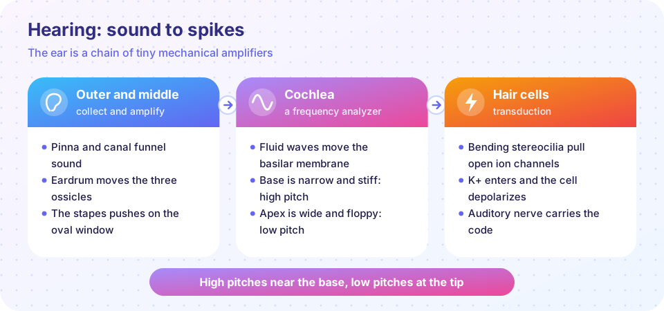
Humans hear roughly from 20 Hz to 20,000 Hz, with the best sensitivity near 1 to 4 kHz. The pinna and ear canal funnel sound to the eardrum. Three tiny bones, the malleus, incus and stapes (the ossicles), carry its motion to the oval window, the membrane at the entrance to the cochlea.
Why bones? Air is easy to compress, fluid is not, and most sound energy would reflect off a water surface. The middle ear is an impedance matcher: the eardrum is much larger than the oval window (about 17 times in area), and the ossicles act as a small lever. Together they raise the pressure by roughly 20-fold. Two tiny muscles can stiffen the chain in loud sound, a protective reflex.
The cochlea: a frequency analyzer
The cochlea is a coiled tube divided lengthwise by the basilar membrane. Pressure waves entering at the oval window travel along it. The membrane is narrow and stiff at the base (near the oval window) and wide and floppy at the apex. As a result high frequencies peak near the base and low frequencies near the apex. Georg von Békésy measured this traveling wave in cadavers and received the 1961 Nobel Prize.
Sitting on the basilar membrane is the organ of Corti. It holds about 3,500 inner hair cells, which are the main sensory cells, and about 12,000 outer hair cells, which amplify. Outer hair cells change their length with voltage (they use a motor protein called prestin) and add energy to the vibration, sharpening the tuning by a large factor. Damage to hair cells, from loud noise, drugs or aging, causes permanent hearing loss because human hair cells do not regenerate.
Transduction
Each hair cell has a bundle of stereocilia, ordered by height and joined by fine "tip links". When the basilar membrane moves, the bundle bends. Tension on the tip links opens mechanically gated channels. The fluid bathing the tips (endolymph) is unusually rich in potassium and held at a positive potential, so K⁺ flows in and depolarizes the cell. Calcium channels open, the cell releases glutamate onto auditory nerve fibers, and spikes carry the signal to the brain. The whole process is fast enough to follow the phase of sound waves up to a few kilohertz.
Central pathway and localization
Auditory nerve fibers synapse in the cochlear nuclei of the brainstem. Signals from both ears meet in the superior olive, where two cues for horizontal location are computed:
- Interaural time difference (ITD): a sound from the left reaches the left ear a fraction of a millisecond earlier. The largest ITD for a human head is about 0.6 to 0.7 ms.
- Interaural level difference (ILD): the head casts a "shadow", so high-frequency sounds are quieter at the far ear.
Pathways continue through the inferior colliculus and the medial geniculate nucleus of the thalamus to the primary auditory cortex in the temporal lobe, where frequency is again mapped topographically.
Balance
Next to the cochlea sit the vestibular organs. Three semicircular canals at right angles sense head rotation: fluid lags when the head turns and bends hair cells. The utricle and saccule contain crystals (otoliths) on a gel; they sense linear acceleration and the pull of gravity. The vestibulo-ocular reflex uses this signal to move the eyes opposite to head rotation, keeping the image stable when you walk or nod. Dizziness often comes from a mismatch between vestibular, visual and body-position signals.
Common misconceptions
"Pitch and loudness are coded the same way." Place along the basilar membrane (and spike timing at low frequencies) codes pitch; firing rate and the number of active fibers code intensity.
"Hearing loss can be reversed by resting the ears." Temporary threshold shifts do recover, but repeated loud exposure can destroy hair cells permanently.
Try it
Press Run (or Ctrl + Enter) to see the result here.
Program input · what you'd type while the program runs
Type the answers before you press Run, one per line. Each time the program reads a line (with input()), it gets the next line from this box.
Press Run (or Ctrl + Enter) to see the result here.
Program input · what you'd type while the program runs
Type the answers before you press Run, one per line. Each time the program reads a line (with input()), it gets the next line from this box.
Check your understanding
Quick check · 5 questions
of 5 answered You scored / 5 · perfect!
-
1. Why does the middle ear exist?
Area ratio and the ossicle lever increase pressure so sound is not mostly reflected at the fluid boundary. -
2. Where on the basilar membrane do high frequencies peak?
The base is narrow and stiff, so it vibrates best at high frequencies. -
3. What ion mainly enters a hair cell to depolarize it?
Endolymph is rich in K+ and positively charged, so K+ flows in when channels open. -
4. The interaural time difference is used to judge what?
A tiny difference in arrival time between ears signals the side of the source. -
5. Which structure keeps the eyes steady when you turn your head?
Semicircular canal signals drive eye movements opposite to head rotation.
Summary
- The outer and middle ear collect sound and increase pressure for the fluid-filled cochlea.
- The basilar membrane maps frequency to place; outer hair cells amplify.
- Bending stereocilia open channels and K⁺ depolarizes inner hair cells.
- The superior olive compares timing and level between ears to locate sounds.
- Semicircular canals and otoliths sense head rotation and linear acceleration.
Track your progress
Create a free account to tick off lessons and pick up where you left off on any device.
of Neuroscience lessons completed