


















Lesson 38 of 45 · Disease and Neurotechnology
Windows into the living brain: neurotechnology
Compare EEG, MEG, fMRI, calcium imaging and optogenetics, and learn how deep brain stimulation and brain-computer interfaces treat and decode the brain.
A living brain is hard to study: it is enclosed in bone, electrically noisy, and made of billions of cells. Every measurement technique trades space against time and invasiveness against detail. Knowing the trade-offs lets you read a news headline about a "brain scan" critically and choose the right tool for a question.
What you will learn
- What EEG and fMRI measure and their limits
- How optogenetics and imaging work in animals
- How deep brain stimulation and brain-computer interfaces help patients
- The ethical questions of reading and changing brains
Key terms
EEG: electroencephalography, scalp voltage recordings.
BOLD signal: blood-oxygen-level-dependent signal measured by fMRI.
Optogenetics: controlling neurons with light by expressing light-sensitive proteins.
DBS: deep brain stimulation through implanted electrodes.
BCI: a brain-computer interface that turns neural activity into commands.
Recording brain activity
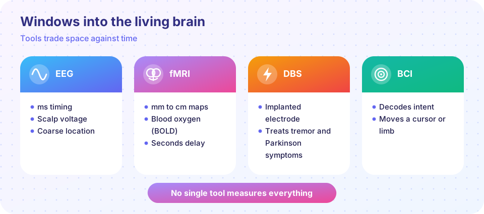
EEG. In 1924 Hans Berger recorded the first human EEG. Electrodes on the scalp pick up summed voltage fluctuations from large groups of aligned cortical neurons. EEG has millisecond timing and is cheap and portable, but it blurs location because bone and tissue smear the signal. Rhythms are named by frequency: delta (0.5 to 4 Hz, deep sleep), theta (4 to 8 Hz), alpha (8 to 13 Hz, relaxed with eyes closed), beta (13 to 30 Hz, active thinking) and gamma (above about 30 Hz). Event-related potentials average many trials to reveal responses to a stimulus. MEG records the tiny magnetic fields instead and localizes better.
fMRI. Functional magnetic resonance imaging does not measure neurons directly. Active neurons need oxygen, local blood flow rises more than needed, and the change in oxygenated hemoglobin alters the MRI signal. This is the BOLD signal (Seiji Ogawa, 1990). Spatial resolution is millimeters and whole-brain coverage is possible, but the hemodynamic response is slow: it rises over a few seconds and peaks around 5 s after the neural event. fMRI maps where regions respond; it cannot easily say how. Statistical care matters: an infamous 2009 study showed "brain activity" in a dead salmon when corrections for multiple comparisons were skipped.
Single cells and imaging. In animals, microelectrodes record single neurons, and modern silicon probes (such as Neuropixels) record hundreds at once. Two-photon calcium imaging uses fluorescent proteins whose brightness rises with calcium, so individual neurons' activity is visible through a window in the skull. In human patients, electrodes placed on or in the brain for epilepsy surgery sometimes provide rare direct recordings.
Changing brain activity
TMS (transcranial magnetic stimulation, Barker 1985) uses a magnetic pulse to induce current in cortex without surgery; repetitive TMS is approved for some treatment-resistant depression. Optogenetics (Karl Deisseroth, Edward Boyden, Gero Miesenböck and others, from about 2005) places light-gated channels such as channelrhodopsin in chosen cell types, so light can turn those neurons on or off with millisecond precision. It is a powerful research tool in animals and is only starting in human trials, for example in vision restoration.
Deep brain stimulation. Surgeons implant an electrode, wired to a pulse generator under the skin of the chest, in a target such as the subthalamic nucleus. High-frequency stimulation (about 130 Hz) reduces tremor, rigidity and slowness in selected patients with Parkinson disease and treats essential tremor and dystonia. Why it works is still debated: it may silence, excite or reshape pathological rhythms. DBS is also studied for depression and obsessive-compulsive disorder.
Brain-computer interfaces. Recall the population vector from the motor cortex lesson. Implanted electrode arrays in people with paralysis let them control a cursor or a robotic arm by thinking about movement (BrainGate and other groups). Since the early 2020s, teams have decoded attempted speech from motor cortex activity into text at rates many times faster than earlier devices, and have used stimulation of sensory cortex to create touch sensations. Cochlear implants, which stimulate the auditory nerve, are the most widely used neural prosthesis, in more than a million people worldwide.
Ethics
Neural data are intimate. Questions include privacy and consent for brain data, security of implants, fair access, the risk of overselling (brain scans cannot read minds reliably), and identity when stimulation changes mood. Researchers and some countries have begun to discuss "neurorights".
Common misconceptions
"fMRI shows neurons firing in real time." It shows blood-oxygen changes seconds after activity.
"Bright regions in a scan are the 'center' of a function." Most functions involve networks, and activity is not the same as necessity.
"EEG can read thoughts." It can detect broad states and some patterns; it does not decode private thought.
Try it
Press Run (or Ctrl + Enter) to see the result here.
Program input · what you'd type while the program runs
Type the answers before you press Run, one per line. Each time the program reads a line (with input()), it gets the next line from this box.
Press Run (or Ctrl + Enter) to see the result here.
Program input · what you'd type while the program runs
Type the answers before you press Run, one per line. Each time the program reads a line (with input()), it gets the next line from this box.
Check your understanding
Quick check · 5 questions
of 5 answered You scored / 5 · perfect!
-
1. Which tool has millisecond timing but coarse spatial resolution?
EEG follows fast voltage changes but blurs where they originate. -
2. What does the BOLD signal reflect?
fMRI measures hemodynamic changes that follow activity by seconds. -
3. What does optogenetics let researchers do?
Light-gated channels are expressed in chosen cell types. -
4. DBS for Parkinson disease commonly targets which structure?
The STN and GPi are common targets. -
5. Which EEG rhythm is typical of relaxed wakefulness with eyes closed?
Alpha increases over occipital regions when the eyes are closed.
Summary
- EEG and MEG give speed; fMRI gives whole-brain maps but is slow and indirect.
- Animal tools (electrode arrays, calcium imaging, optogenetics) reach cellular detail.
- DBS and BCIs treat and decode; cochlear implants are widely used neuroprostheses.
- Neural data raise privacy and consent issues.
Next: Chapter 8 review
Track your progress
Create a free account to tick off lessons and pick up where you left off on any device.
of Neuroscience lessons completed