


















Lesson 18 of 43 · Neuroscience Foundations
Brain organization and localization
Tour the four lobes and key structures of the brain, famous patient cases such as Phineas Gage and H.M., and the debate between localized and distributed function.
Does one part of the brain do one job? Nineteenth-century physicians began to think so after patients with damage to a particular place lost a particular ability. A century and a half later, imaging shows that the answer is "partly yes, partly no": regions have specialties, but real tasks recruit networks. This lesson surveys the large-scale map of the brain and the famous cases that taught us how to read it.
What you will learn
- The names, positions and broad functions of the four lobes and some deeper structures.
- What the cases of Phineas Gage, Broca's patient, Wernicke's patients and H.M. showed.
- How split-brain studies and double dissociations shaped the field.
- Why modern researchers talk about networks, not single regions.
Key terms
Cerebral cortex: the folded outer sheet of the cerebrum, a few millimeters thick.
Lobe: one of the four major divisions of each cerebral hemisphere (frontal, parietal, temporal, occipital).
Localization of function: the idea that specific functions depend on specific regions.
Lesion: damage to brain tissue.
Anterograde amnesia: inability to form new long-term memories after the damage.
The big map
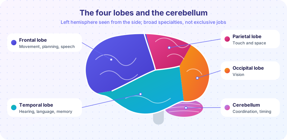
The cerebrum has two hemispheres, joined mainly by a thick bundle of fibers, the corpus callosum. Each hemisphere is divided into four lobes, named after the skull bones above them.
| Lobe | Position | Broad functions |
|---|---|---|
| Frontal | Front, ahead of the central sulcus | Movement (primary motor cortex), planning, working memory, decision making, speech production |
| Parietal | Top and back, behind the central sulcus | Touch and body sense (primary somatosensory cortex), spatial attention, integrating senses |
| Temporal | Side, below the lateral sulcus | Hearing, language comprehension, object recognition, long-term memory (medial parts) |
| Occipital | Back | Vision (primary visual cortex) |
Beneath and behind the cortex lie structures with their own jobs: the thalamus (relay for most sensory signals), the hippocampus (forming new memories), the amygdala (emotional significance), the basal ganglia (action selection and habit), the cerebellum (coordination and timing of movement, with growing evidence for roles in cognition) and the brainstem (breathing, heart rate, arousal).
Around 1909 Korbinian Brodmann divided the cortex into about 52 areas by how the cells look under a microscope. "Area 17" (primary visual cortex) and "Area 4" (primary motor cortex) are still used as names. Wilder Penfield's stimulation studies on surgical patients in the mid-twentieth century revealed that the motor and somatosensory strips contain a distorted body map, the "homunculus", with large areas for the hands and mouth.
Famous cases
Phineas Gage (1848)
A railroad foreman, Gage survived an explosion that drove an iron rod through the front of his skull. His doctor, John Harlow, reported that his personality changed. Gage's case is often cited as showing that the frontal lobes help regulate social behavior and planning. The popular story is exaggerated: contemporary accounts are sparse, and later evidence suggests that Gage regained considerable function and held a job for years. The case is a good reminder to check sources.
Broca's patient (1861)
Paul Broca examined a man known as "Tan", the only syllable he could say, though he understood speech. After the man died, Broca found damage in the left frontal lobe. Over time he collected similar cases. Broca's area is associated with speech production, and damage causes slow, effortful, grammatically reduced speech with relatively preserved comprehension.
Wernicke's patients (1874)
Carl Wernicke described patients with fluent, well-formed speech that made little sense, and poor comprehension, after damage to the left posterior temporal lobe. This suggested separate regions for producing and understanding language. The classical model linked them with a fiber tract. Modern research finds language to be more widely distributed, but the contrast between the two syndromes remains a basic clinical distinction.
H.M. (1953)
To treat severe epilepsy, surgeon William Scoville removed large portions of the medial temporal lobes of Henry Molaison (identified as H.M. until his death in 2008). His epilepsy improved, but he could no longer form new long-term memories of facts and events, while keeping his intelligence, language and short-term memory. Brenda Milner showed that he could still learn new motor skills, such as mirror drawing, without remembering practicing them. This revealed that memory is not one thing: the hippocampal region is crucial for new declarative memories, while skill learning depends on other systems.
Split-brain patients
To limit the spread of seizures, surgeons cut the corpus callosum in some patients. Roger Sperry and Michael Gazzaniga then showed that the two hemispheres could be tested separately, using images flashed to one side of the visual field. The left hemisphere typically could name what it saw, while the right hemisphere could pick out objects with the left hand without being able to say what they were. Sperry received the Nobel Prize in 1981. The popular conclusion that people are "left-brained" or "right-brained" is a large overstatement of these results.
Localization versus distribution
The early cases support localization. But in rat experiments, Karl Lashley (1929) found that the amount of cortex removed mattered more than its location for maze learning, which he described as "mass action" and "equipotentiality". Both views contain truth. Primary sensory and motor areas are clearly specialized. Higher functions such as language, memory and attention involve networks of regions that work together, and a region may take part in many networks.
Researchers use dissociations to infer separable mechanisms. A single dissociation (a patient impaired on A but not B) can arise because A is simply harder. A double dissociation (one patient with the reverse pattern) is stronger evidence for separate processes. The idea of a double dissociation was developed by Hans-Lukas Teuber in the 1950s.
Common misconceptions
"Each brain region has exactly one function." Most regions participate in many tasks, and most tasks involve several regions.
"Phineas Gage became a different person overnight and stayed that way." Reports were limited and later evidence points to partial recovery.
"Broca's and Wernicke's areas are the language centers." They are important, but language relies on a broad network of regions in both hemispheres, with the left usually dominant.
Try it
A lobe explorer. Click a lobe to see its main roles. The map is simplified, and the boundaries are approximate.
A double-dissociation detector. Given hypothetical patient scores on two tasks, the code flags impaired performance (more than 2 standard deviations below the control mean) and looks for the opposite patterns in two patients. All data are invented for the exercise.
Press Run (or Ctrl + Enter) to see the result here.
Program input · what you'd type while the program runs
Type the answers before you press Run, one per line. Each time the program reads a line (with input()), it gets the next line from this box.
Check your understanding
Quick check · 5 questions
of 5 answered You scored / 5 · perfect!
-
1. Which lobe contains primary visual cortex?
The occipital lobe, at the back of the brain, houses primary visual cortex. -
2. What did the case of H.M. reveal?
H.M. could not form new memories of events but could still learn motor skills. -
3. Broca's area damage typically causes
Broca-type aphasia mainly affects production. -
4. Why is a double dissociation stronger evidence than a single dissociation?
The reversed pattern in a second patient rules out the explanation that one task is just harder. -
5. Which statement about the Phineas Gage story is best supported?
Historical records are limited, and evidence points to partial recovery.
Summary
- The cortex has four lobes (frontal, parietal, temporal, occipital) with broad specialties, plus deeper structures such as the hippocampus, thalamus and cerebellum.
- Cases such as Broca's patient, Wernicke's patients and H.M. showed that damage to particular regions can selectively impair functions.
- Split-brain studies revealed hemispheric differences but do not support the "left-brain, right-brain" personality myth.
- Modern views stress networks and use double dissociations to infer separable processes.
Track your progress
Create a free account to tick off lessons and pick up where you left off on any device.
of Cognitive Science lessons completed