


















Lesson 37 of 49 · Structural Bioinformatics
Structure Prediction and Molecular Docking
Learn how protein structures are predicted from sequence, how to read AlphaFold's pLDDT and PAE confidence, and how docking scores ligands, with limits and pitfalls.
Getting a protein's structure used to require months or years in a laboratory. Today, for most proteins, you can type a sequence into a program and get a 3D model in minutes, or download one from a public database. This changed biology more quickly than almost anything else in the last decade. It also created a new skill: knowing how much to trust a predicted model, and what questions it can and cannot answer.
This lesson explains how prediction works, how to read the confidence numbers that come with a model, and how docking uses a structure to estimate how a small molecule might bind. The two problems are linked: a good pocket in a predicted model is the starting point for a docking study, and both are computational estimates that need laboratory confirmation.
What you will learn
- The protein folding problem and the main families of prediction methods.
- How AlphaFold and similar tools work in outline, and what pLDDT and PAE mean.
- What docking does: pose search plus scoring.
- The most important limits of both approaches and how to avoid over-reading results.
Key terms
Homology modelling: building a model of a protein from the known structure of a related protein (the template).
Multiple sequence alignment (MSA): an alignment of many related sequences, used to find conserved and co-evolving positions.
pLDDT: predicted local distance difference test, a per-residue confidence from 0 to 100.
PAE (predicted aligned error): the expected positional error between two residues, shown as a matrix.
Pose: the position, orientation and conformation of a ligand in a binding site.
The folding problem
In 1973 Christian Anfinsen showed that a protein's sequence contains the information needed to specify its fold: unfolded ribonuclease refolded on its own into the active enzyme. That implies that the structure, in principle, can be computed from the sequence. But the number of possible conformations of a chain is astronomically large (the point of Levinthal's paradox: a protein cannot find its fold by random search, so folding must be guided). For fifty years, predicting a fold from sequence alone stayed one of biology's grand challenges. Since 1994 the CASP experiment (Critical Assessment of Structure Prediction) has tested methods every two years on structures solved but not yet released, which kept the field honest.
Families of methods
Homology (comparative) modelling. Proteins with similar sequences share similar folds. If a related protein of known structure exists (the template, typically with sequence identity above roughly 30%), you align the target to it, copy the backbone, model the loops and side chains, refine and evaluate. Tools include MODELLER and the SWISS-MODEL web service. Accuracy falls as identity falls.
Fragment assembly and physics-guided methods. Rosetta, from David Baker's group, assembles structures from short fragments of known proteins guided by an energy function. It also underlies computational protein design.
Co-evolution and deep learning. Look at an alignment of thousands of related sequences. If two positions far apart in the sequence tend to mutate together, they are probably in contact in 3D, because a change at one must be compensated by a change at the other. Early methods extracted such contacts statistically. In 2018 DeepMind's AlphaFold used deep learning on them at CASP13. In 2020 AlphaFold 2 reached accuracy comparable to experiments for most CASP14 targets (a median backbone error of roughly 1 Å), and its method was published in 2021. It processes the MSA and a pairwise representation of residues with attention-based networks (the Evoformer), then a structure module that outputs 3D coordinates, refined by recycling the output. RoseTTAFold (Baker lab, 2021) follows a similar idea, ESMFold (Meta, 2022) predicts from a single sequence using a protein language model, and AlphaFold 3 (2024) extends prediction to complexes including nucleic acids and small molecules. The 2024 Nobel Prize in Chemistry recognized this work: half to David Baker for computational protein design, and half to Demis Hassabis and John Jumper for AlphaFold.
The AlphaFold Protein Structure Database, launched in 2021 with EMBL-EBI, holds predicted models for over 200 million sequences, so for many proteins you do not run anything; you simply download the file.
Reading the confidence information
A predicted model is useless without its uncertainty. Two outputs matter.
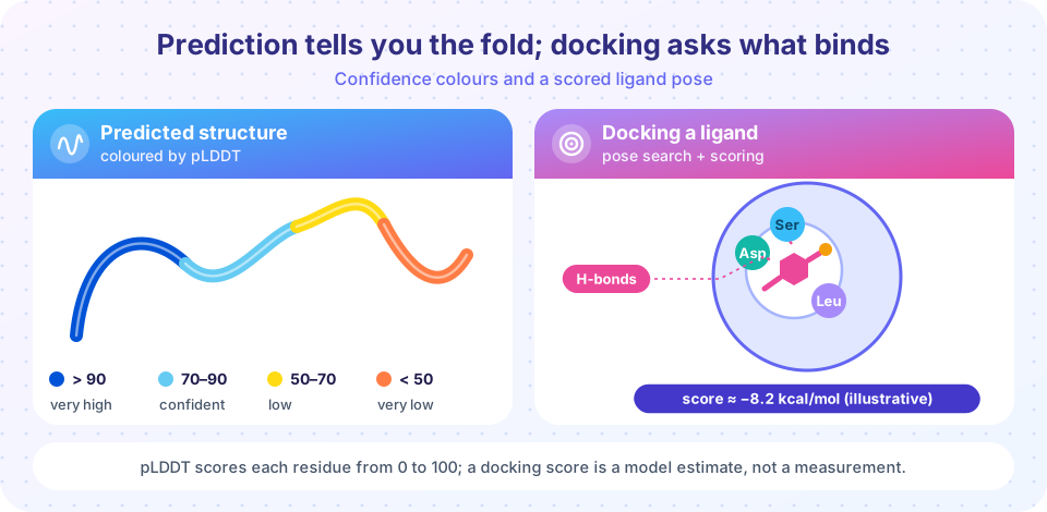
pLDDT gives each residue a score from 0 to 100, stored in the B-factor column of the model file (not a temperature factor in this case). The conventional bands, matching the figure:
| pLDDT | Interpretation | Colour in viewers |
|---|---|---|
| above 90 | very high: backbone and often side chains reliable | dark blue |
| 70 to 90 | confident: backbone usually good | light blue |
| 50 to 70 | low: treat with caution | yellow |
| below 50 | very low: often intrinsically disordered, shown as a spaghetti-like tail | orange |
Low scores are informative, not just a failure: very low pLDDT in an isolated stretch is a good sign that the region is disordered in reality.
PAE is a matrix with a value for each pair of residues: if you aligned the structure on residue A, how far off would residue B be? Low PAE between two domains means their relative arrangement is trustworthy; high PAE means each domain may be right but how they pack together is not. For complexes, scores like ipTM estimate interface quality.
Limits to remember
- A model is one conformation. Proteins move, switch states and bind partners, and a single prediction does not show that.
- Effects of point mutations are not reliably captured: a model of a mutant often looks like the wild type.
- Cofactors, metal ions, post-translational modifications and ligands are absent or only partially handled in many tools.
- Proteins with few homologs have shallow alignments and lower accuracy.
- A confident model can still be wrong in the details that matter (an active-site side chain), so validate with experiments where decisions depend on it.
Molecular docking
Docking predicts how a small molecule (a ligand) binds to a protein. It has two parts that work together.
- Search. Generate many candidate poses by moving and rotating the ligand and twisting its flexible bonds inside a search box around the pocket. Methods include genetic algorithms, Monte Carlo sampling with local optimization (AutoDock Vina) and systematic fragment growth (Glide, GOLD). Recent diffusion models such as DiffDock generate poses by learned denoising.
- Scoring. Assign each pose a number that estimates how favorable it is. Force-field scores sum van der Waals and electrostatic terms; empirical scores fit weights to terms like hydrogen bonds, hydrophobic contact and the loss of ligand flexibility; knowledge-based scores come from contact statistics in the PDB; machine-learned scores are trained on affinity data. Scores are usually reported in kcal/mol, with more negative meaning better.
The most basic building block is the Lennard-Jones potential for a pair of atoms at distance r:
V(r) = 4 * epsilon * ((sigma / r)^12 - (sigma / r)^6)
The r^-12 term is a steep repulsion at short range (atoms cannot overlap), the r^-6 term is a weak attraction at medium range, and the minimum lies at r = 2^(1/6) * sigma with depth -epsilon. Real scoring functions add hydrogen-bond, electrostatic and desolvation terms on top.
A typical virtual screening workflow: prepare the receptor (add hydrogens, assign protonation states, remove or keep key waters), prepare a library of ligands in 3D, define the search box, dock all of them, rank by score, inspect the top poses by eye for sensible chemistry, optionally refine with molecular dynamics, then test the best candidates in an assay. A docking study is a way to prioritize experiments, not to replace them.
How docking is validated
- Redocking: dock a ligand back into its own crystal structure and measure the RMSD of the best pose to the experimental one (see the RMSD lesson); below about 2 Å is usually called a success.
- Enrichment: mix known actives with decoy molecules and check that actives rank near the top.
Pitfalls
- Scores correlate only loosely with measured binding affinities, so do not read small score differences as real.
- Most programs treat the receptor as rigid, though proteins reshape on binding.
- Ordered water molecules, protonation and tautomer states can change the result.
- A pose can look beautiful and still be wrong; the search may have fallen into a local optimum.
This lesson describes computational methods for education. It is not advice about any drug, treatment or medical decision.
Common misconceptions
"AlphaFold solved protein folding." It predicts native-like structures for most single, well-folding proteins with impressive accuracy, but it does not explain the folding process, protein dynamics or the effect of mutations.
"High pLDDT means the structure is certainly correct everywhere." It is a confidence estimate for local geometry. Relative domain positions need PAE, and function-critical details still need experiments.
"A better docking score means a better drug." The score is a rough model of binding. Selectivity, solubility, toxicity and many other properties decide whether a molecule becomes a drug.
Try it
First, analyze a per-residue confidence profile the way you would for a real AlphaFold file, finding the domains and the disordered stretches. Second, run a toy 2D docking search with a Lennard-Jones plus hydrogen-bond score (a teaching simulation, not a real docking program). Third, explore the Lennard-Jones curve interactively.
Press Run (or Ctrl + Enter) to see the result here.
Program input · what you'd type while the program runs
Type the answers before you press Run, one per line. Each time the program reads a line (with input()), it gets the next line from this box.
Press Run (or Ctrl + Enter) to see the result here.
Program input · what you'd type while the program runs
Type the answers before you press Run, one per line. Each time the program reads a line (with input()), it gets the next line from this box.
Check your understanding
Quick check · 5 questions
of 5 answered You scored / 5 · perfect!
-
1. In an AlphaFold model, a stretch of 25 residues has pLDDT below 40. The best interpretation is:
Very low pLDDT often corresponds to intrinsically disordered regions and means the shown coordinates are unreliable. -
2. Which output would you check to judge how two domains are arranged relative to each other?
PAE reports expected positional error between residue pairs, so it reveals whether domain packing is trustworthy. -
3. A docked pose of a ligand has an RMSD of 1.4 Å to the crystal pose in a redocking test. How is this usually judged?
Redocking success is conventionally an RMSD below about 2 Å. -
4. The Lennard-Jones potential has its minimum energy at which distance?
Setting the derivative to zero gives r = 2^(1/6) * sigma, where V = -epsilon. -
5. Which is a known weakness of single-structure predictions?
Proteins are dynamic, and a prediction usually gives one state; mutation effects are not reliably captured.
Summary
- Prediction methods range from homology modelling to co-evolution-based deep learning (AlphaFold 2 and 3, RoseTTAFold, ESMFold); models are freely available for hundreds of millions of sequences.
- Use pLDDT (stored in the B-factor column) for per-residue confidence and PAE for relative domain placement; low pLDDT often flags disorder.
- Models are single conformations; they are weak on mutation effects, ligands and shallow alignments.
- Docking combines pose search with a scoring function (Lennard-Jones, hydrogen bonds, desolvation and more); validate by redocking (under about 2 Å) and enrichment.
- Scores rank ideas, they do not prove binding; confirm in the laboratory.
Track your progress
Create a free account to tick off lessons and pick up where you left off on any device.
of Bioinformatics lessons completed