


















Lesson 38 of 49 · Structural Bioinformatics
Chapter 7 Review: Structural Bioinformatics
Review protein structure levels, PDB coordinates, Ramachandran angles, superposition and RMSD, AlphaFold confidence and docking, with a recap table and a ten-question quiz.
This chapter moved from strings and counts to coordinates. You learned that a protein is described at four nested levels, that a PDB file is a table of atoms with fixed columns, that geometry (distances and dihedral angles) turns that table into knowledge, that structures are compared by superposition and RMSD, and that predicted models and docked poses come with confidence measures you must read before you believe them.
Use this review to check that those ideas hang together, and to practice the habit that matters most in structural work: asking "how good is this model, and for which question?" before drawing conclusions.
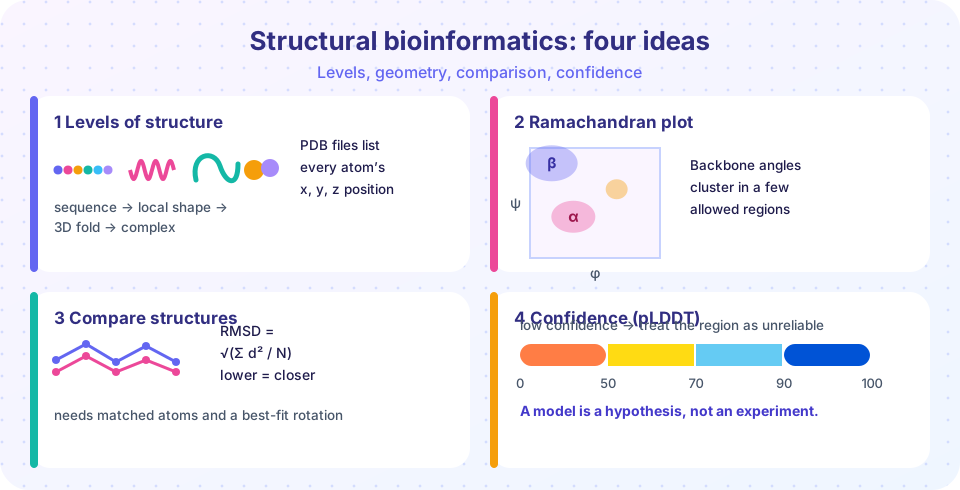
The chapter in one table
| Idea | Key facts | Lesson |
|---|---|---|
| Levels of structure | primary, secondary (helix, sheet), tertiary, quaternary; resolution in ångströms, lower is better | Overview |
| PDB files | fixed-column ATOM lines; x, y, z in Å; occupancy; B-factor; mmCIF for large structures | PDB files |
| Local geometry | consecutive C-alpha atoms 3.8 Å apart; phi and psi; helix 3.6 residues per turn, i to i+4 hydrogen bonds | PDB files |
| Comparison | RMSD = sqrt(mean of d^2) after Kabsch superposition; TM-score, GDT_TS, lDDT |
Superposition |
| Prediction | homology modelling, co-evolution and deep learning; pLDDT 0 to 100; PAE for domain placement | Prediction and docking |
| Docking | pose search plus scoring; Lennard-Jones, hydrogen bonds; redocking under about 2 Å | Prediction and docking |
Ideas that connect the lessons
Coordinates are a model, with uncertainty attached. B-factors, occupancy and resolution for experimental structures, and pLDDT and PAE for predicted ones, are all the same kind of information: how much each part of the model can be trusted. In AlphaFold files, pLDDT even sits in the B-factor column, so the two ideas literally share a field.
Distances are the universal language. Contact maps, RMSD, lDDT, the 3.8 Å Cα spacing, hydrogen-bond lengths and the Lennard-Jones minimum are all statements about distances between atoms. If you can compute a distance matrix, you can do a surprising amount of structural analysis without any specialized software.
Rigid-body alignment is a modelling choice. Superposition assumes parts of the molecule move together. When that fails (hinges, flexible loops), global RMSD stops being meaningful, which is why domain-wise comparison and superposition-free measures exist.
Sampling and scoring are separate problems. In docking, the search can find the correct pose while the scoring function ranks it poorly, or vice versa. When a result disappoints, ask which half failed.
A reproducible checklist
Before using a structure for any conclusion:
- Source and method: experimental (X-ray, NMR, cryo-EM) or predicted? Which resolution or which model version?
- Local quality: B-factors, occupancy and missing residues near the region of interest; pLDDT and PAE for predicted models.
- Geometry: no chain breaks (Cα spacing near 3.8 Å), no serious clashes, few Ramachandran outliers.
- Comparison: state which atoms, which alignment and which superposition produced the RMSD or TM-score.
- Docking claims: report the validation (redocking, enrichment), the receptor preparation and the software version.
Worked example: Is this model good enough for docking?
A collaborator proposes docking a library of molecules into an AlphaFold model of an enzyme from a poorly studied bacterium. How do you decide whether to proceed?
Start with the confidence data. The overall mean pLDDT is not enough; look at the residues lining the proposed active site. If most are above 90, the backbone around the pocket is probably accurate, but side-chain rotamers can still differ from the real enzyme. Next, check whether the enzyme normally binds a cofactor or metal ion: many predicted models lack them, which changes the shape and charge of the pocket. Third, ask whether the enzyme changes shape on binding; if it closes over its substrate, a rigid receptor docking study will miss the relevant state.
Then run a sanity check. If a homologous enzyme has an experimental structure with a bound inhibitor, superpose the model on it and measure the RMSD of the pocket residues. Redock the known inhibitor into the model; if the best pose is more than 2 Å from the experimental one, the model (or the protocol) is not good enough yet. Only if these checks pass does docking a library make sense, and even then the output is a list of ideas to test at the bench, not a list of results.
Common slips to avoid
Structural analyses go wrong in predictable ways, and naming the slips makes them easier to catch.
Mixing up numbering schemes. A PDB file may number residues differently from the UniProt sequence, because of expression tags, missing segments or engineered constructs. Before comparing residue 120 in one structure with residue 120 in another, check that they are the same amino acid in the same protein. Comparing unequal atom sets. RMSD over all heavy atoms and RMSD over C-alpha atoms are different numbers; report which you used. Forgetting alternate conformations and missing density. Atoms with partial occupancy, or residues with no coordinates at all, can quietly change a distance or a contact count. Trusting hydrogens that were not measured. X-ray structures rarely show hydrogens, so hydrogen-bond analyses depend on assumptions about protonation. Reading a pretty picture as evidence. A cartoon drawn in a viewer looks equally confident whether it came from a 1 Å crystal or a low-confidence prediction, so always check the numbers behind the colors.
If you build the habit of checking numbering, atom sets, occupancy and confidence before every measurement, you will avoid the majority of avoidable errors in this field.
Which tool for which job?
Structural questions tend to arrive in a few standard shapes, and each has a standard first move.
"What does this protein look like?" Search the PDB by name or sequence first. If an experimental structure exists, read its resolution and the quality information before looking at the picture. If none exists, look in the AlphaFold database, and read the pLDDT coloring before the ribbon.
"Did this mutation, drug or condition change the shape?" Superpose the two structures on a stable core, then plot per-residue distances. A single RMSD hides whether one loop moved a lot or everything moved a little. Report the atoms, the alignment and the cut-off.
"Where could a small molecule bind?" Look for a pocket (a cavity lined with residues that fit hydrogen bonds and hydrophobic contacts), check whether known ligands bind related proteins, and only then dock. Treat docking scores as a ranking aid and finish with an independent assay.
"Are these two proteins related?" Compare sequences first, since fast alignment is cheap. If the sequences have diverged beyond recognition, compare structures with a tool such as TM-align or Foldseek, because folds are conserved over longer evolutionary distances than sequences.
Notice what the four situations have in common: each starts by checking how reliable the data are and ends by asking what an independent line of evidence would show. That habit is the real lesson of this chapter.
Try it
The first example is a structure sanity checker that flags chain breaks and atomic clashes in a Cα trace. The second is a Ramachandran region classifier you can drive with two sliders.
Press Run (or Ctrl + Enter) to see the result here.
Program input · what you'd type while the program runs
Type the answers before you press Run, one per line. Each time the program reads a line (with input()), it gets the next line from this box.
Check your understanding
Quick check · 9 questions
of 9 answered You scored / 9 · perfect!
-
1. Consecutive C-alpha atoms in a chain are about how far apart?
About 3.8 Å for trans peptides, a quick check for chain breaks. -
2. Which pair of angles defines the local conformation of a protein backbone residue?
Phi and psi describe rotations about the N-C-alpha and C-alpha-C bonds. -
3. Which of the following is NOT a measure of structural similarity?
Phred scores describe sequencing base-call quality; the others compare structures. -
4. Three matched pairs are 2 Å, 2 Å and 2 Å apart after superposition. The RMSD is:
sqrt((4 + 4 + 4) / 3) = sqrt(4) = 2 Å. -
5. In AlphaFold model files, which column stores the pLDDT value?
pLDDT is written in the B-factor field so viewers can color by confidence. -
6. What is the main reason to superpose domains separately for a hinge protein?
Global RMSD becomes large and uninformative when the domains swing relative to each other. -
7. A docking program places the ligand 3.4 Å RMSD from its crystal pose in a redocking test. What does that suggest?
Redocking is conventionally judged successful below about 2 Å. -
8. Which interaction distance is typical for a hydrogen bond between donor and acceptor heavy atoms?
Hydrogen bonds between donor and acceptor atoms are typically about 2.6 to 3.2 Å long. -
9. A residue has phi = -62 and psi = -41. It most likely sits in:
Those angles are the classic right-handed alpha-helix region.
Summary
- Four levels of structure, from sequence to complexes; structures are models with resolution, B-factors and occupancy.
- Geometry from coordinates: distances, 3.8 Å Cα spacing, phi/psi and Ramachandran regions, helices at 3.6 residues per turn.
- Compare structures with Kabsch superposition and RMSD, and complement it with TM-score, GDT_TS and lDDT for hinge or length problems.
- Predicted models carry pLDDT and PAE; docking separates sampling from scoring and is validated by redocking and enrichment.
- Always ask how good the model is for the specific question.
Track your progress
Create a free account to tick off lessons and pick up where you left off on any device.
of Bioinformatics lessons completed