


















Lesson 34 of 49 · Structural Bioinformatics
Chapter 7 Overview: Structural Bioinformatics
A map of structural bioinformatics: how protein structures are determined and stored, the four levels of structure, how structures are compared, predicted and docked.
A protein's sequence is a one-dimensional string of amino acids, but the protein does its work as a three-dimensional machine. An enzyme works because a pocket of the right shape and chemistry sits next to the right atoms. A drug works because its shape fits one such pocket. A mutation harms because it disturbs a fold. Structural bioinformatics is the computational study of these shapes: storing them, measuring them, comparing them, predicting them and asking what binds to them.
In this chapter the data change character once more. Instead of strings of letters or tables of counts, you will work with coordinates: lists of x, y, z positions for thousands of atoms. Geometry replaces statistics as the main tool, though both appear.
What you will learn
- How structures are determined experimentally and where they are stored.
- The four levels of protein structure and the interactions that hold a fold together.
- How to read a PDB file and measure distances and angles from coordinates.
- Why comparing structures needs superposition, and what RMSD does and does not tell you.
- What structure prediction and docking can and cannot do.
Key terms
Residue: one amino acid within a chain.
Ångström (Å): 0.1 nanometre, the natural length unit for atoms; a typical covalent bond is 1 to 1.5 Å.
PDB (Protein Data Bank): the global archive of experimentally determined 3D structures of proteins, nucleic acids and complexes.
Fold: the overall three-dimensional arrangement of a protein chain.
Ligand: a small molecule that binds to a protein.
Where do structures come from?
Three experimental methods supply almost all the structures in the archive.
- X-ray crystallography. Protein is coaxed into a crystal, an X-ray beam is diffracted through it, and the pattern is converted into an electron-density map into which an atomic model is built. It dominated the field for decades and gives high resolution, but needs crystals. John Kendrew solved the first protein structure, myoglobin, at low resolution in the late 1950s.
- NMR spectroscopy. For smaller proteins in solution, nuclear magnetic resonance gives distance restraints between atoms. The result is usually an ensemble of models that all satisfy the data, which also shows how flexible parts of the protein are.
- Cryo-electron microscopy. A solution of protein is flash-frozen, imaged in thin ice and thousands of particle images are combined into a 3D reconstruction. A technical revolution in the 2010s brought routine near-atomic resolution, and cryo-EM now handles large complexes and membrane proteins that resisted crystals.
All structures are deposited in the Protein Data Bank (PDB), founded in 1971 with a handful of structures and now holding well over 200,000 entries, each with a four-character identifier such as 1UBQ. The worldwide PDB (wwPDB) partnership curates it, and the files are free. Resolution is quoted in ångströms, and lower is better: around 1 Å shows individual atoms, 2 Å is good, and beyond 3.5 Å you can see the fold but not the side chains clearly.
Four levels of structure
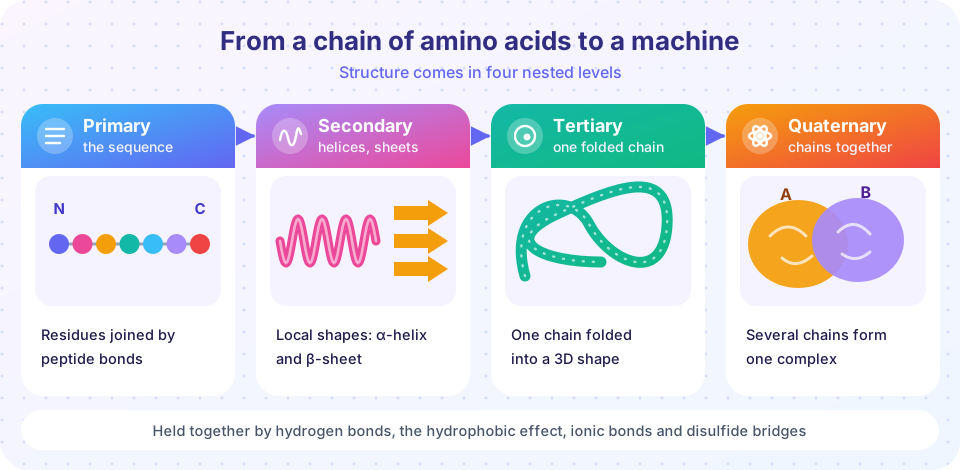
- Primary structure is the sequence of amino acids, joined end to end by peptide bonds. The chain has a direction: from the N-terminus (free amino group) to the C-terminus (free carboxyl group).
- Secondary structure is local repeating shape stabilized by hydrogen bonds between backbone atoms. The two dominant motifs are the alpha helix, a right-handed coil with 3.6 residues per turn, and the beta sheet, made of extended strands side by side. Segments in neither are loosely called coil or loop.
- Tertiary structure is the full three-dimensional fold of a single chain, stabilized by the hydrophobic effect (nonpolar side chains pack into the core), hydrogen bonds, ionic bonds and sometimes disulfide bridges between cysteines.
- Quaternary structure is the arrangement of several chains into one complex, as in hemoglobin's four subunits.
Primary
Set by the gene
Secondary
Held by backbone hydrogen bonds
Tertiary
The unit that does the job
Quaternary
Hemoglobin has four subunits
What structural bioinformatics does with coordinates
Once a structure is a table of coordinates, many questions become computations:
- Measure: distances between atoms, angles along the backbone, the volume of a pocket, the surface exposed to solvent.
- Annotate: assign secondary structure from hydrogen-bond patterns (the DSSP algorithm of Kabsch and Sander, 1983), find ligand-binding sites, list contacts between residues.
- Compare: superpose two structures and quantify how different they are (the RMSD lesson).
- Predict: infer a structure from a sequence (AlphaFold and others), or predict how a small molecule will sit in a pocket (docking).
- Search: find structures with a similar fold, even when the sequences have diverged beyond recognition. Structure is conserved longer than sequence, which is one reason it is so valuable for studying evolution.
Why structure matters beyond curiosity
Structure is where the abstract meets the practical. In medicine, many drugs are small molecules designed to fit a pocket on a protein, and the structure of the target guides which chemical changes might improve the fit. Structures of viral proteins have shown how antibodies recognize them, and the ability to see a protein's shape has shortened the path from a new pathogen's sequence to the design of a vaccine antigen. In genetics, a mutation that changes one amino acid is far easier to interpret when you can see whether the residue sits in the buried core, on a surface that binds a partner, or in a flexible loop.
In biotechnology, enzymes are engineered by changing residues around the active site, and entire proteins that do not exist in nature are now designed from scratch with computational tools. In basic research, the shape of a protein suggests its function even when the sequence looks like nothing known. For all these uses the same discipline applies: a structure is a model with a resolution, a confidence and a context (what was bound, what conditions), and conclusions are only as strong as the part of the model they rest on. The lessons that follow teach you to read the data behind a model before using it.
What each lesson covers
| Lesson | What you will do |
|---|---|
| Protein Structure and PDB Files | Read ATOM records, compute distances and dihedral angles, draw a Ramachandran plot. |
| Superposition and RMSD | Implement the Kabsch algorithm and measure structural differences. |
| Structure Prediction and Docking | Interpret pLDDT confidence, and explore scoring functions and a toy docking search. |
| Chapter Review | Consolidate the four ideas and test them. |
Try it
The first example uses the idea of an ideal helix to generate coordinates and checks a number you can find in any biochemistry text: neighboring alpha carbons are about 3.8 Å apart. The second rotates a helix in 3D with a slider.
Press Run (or Ctrl + Enter) to see the result here.
Program input · what you'd type while the program runs
Type the answers before you press Run, one per line. Each time the program reads a line (with input()), it gets the next line from this box.
Check your understanding
Quick check · 4 questions
of 4 answered You scored / 4 · perfect!
-
1. Which experimental method typically produces an ensemble of models for a small protein in solution?
NMR gives distance restraints, usually satisfied by a bundle of models that also show flexibility. -
2. What forces mainly stabilize alpha helices and beta sheets?
Backbone C=O and N-H groups form the regular hydrogen-bond patterns of secondary structure. -
3. Which level of structure describes the folded 3D shape of a single chain?
Tertiary structure is the full 3D fold of one chain; quaternary is the arrangement of several chains. -
4. A structure has a resolution of 1.2 Å and another has 3.8 Å. Which has finer detail?
A smaller resolution number means finer detail.
Summary
- Structures come from X-ray crystallography, NMR and cryo-EM, and are archived in the PDB, with resolution in ångströms (lower is better).
- Structure has four levels: sequence, local helices and sheets, the folded chain, and multi-chain complexes.
- Structural bioinformatics measures, annotates, compares, predicts and searches coordinates; structure outlasts sequence in evolution.
- The next lessons teach coordinates in practice, superposition and RMSD, and prediction and docking.
Track your progress
Create a free account to tick off lessons and pick up where you left off on any device.
of Bioinformatics lessons completed